Abstract
Purpose
Transforming growth factor-β1 (TGF-β1) plays a dual role in apoptosis and in proapoptotic responses in the support of survival in a variety of cells. The aim of this study was to determine the function of TGF-β1 in bladder cancer cells.
Materials and Methods
The role of TGF-β1 in bladder cancer cells was examined by observing cell viability by using the tetrazolium dye (MTT) assay after treating the bladder cancer cell lines 253J, 5637, T24, J82, HT1197, and HT1376 with TGF-β1. Among these cell lines, the 253J and T24 cell lines were coincubated with TGF-β1 and the pan anti-TGF-β antibody. Fluorescence-activated cell sorter (FACS) analysis was performed to determine the mechanism involved after TGF-β1 treatment in 253J cells.
Results
All six cell lines showed inhibited cellular growth after TGF-β1 treatment. Although the T24 and J82 cell lines also showed inhibited cellular growth, the growth inhibition was less than that observed in the other 4 cell lines. The addition of pan anti-TGF-β antibodies to the culture media restored the growth properties that had been inhibited by TGF-β1. FACS analysis was performed in the 253J cells and the 253J cells with TGF-β1. There were no significant differences in the cell cycle between the two treatments. However, there were more apoptotic cells in the TGF-β1-treated 253J cells.
In human tissues, cells and their surrounding extracellular matrix microenvironment interact throughout all stages of life. These cooperative interactions involve numerous cytokines acting through specific cell-surface receptors. Transforming growth factor β (TGF-β) is a versatile cytokine that is intimately involved in cell growth, differentiation, and immune modulation [1,2,3].
The initial experiments with TGF-β were based on its ability to induce malignant behavior in normal fibroblasts [4]. Such findings resulted in the concept that TGF-β was a key factor in tumorigenesis. However, several years later it emerged that TGF-β has profound growth suppressive effects on many cells, including epithelial cells [5].
TGF-β plays various roles in the process of malignant progression. It is a potent inhibitor of normal stromal, hematopoietic, and epithelial cell growth. However, at some point during cancer development, the majority of transformed cells become either partly or completely resistant to TGF-β growth inhibition [6]. Loss or mutational inactivation of the genes that encode signaling intermediates in the TGF-β pathway have been described to be associated with the resistance to growth inhibition by TGF-β, thus allowing uncontrolled proliferation of the cells [7]. However, the stage of tumor development or progression at which TGF-β-resistant clones come to dominate the tumor cell population in different types of neoplasm remains to be defined.
Bladder cancer is the fourth most common malignancy among western men, following prostate, lung, and colon cancer. In Europe and the United States, bladder cancer accounts for 5% to 10% of all malignancies among males [8]. In Korea, bladder cancer is the second most common urology malignancy following prostate cancer and accounted for 2.7% of all malignancies among males in 2010 [9].
In the present study, we studied the role of TGF-β in bladder cancer cells. These findings can help to establish a treatment strategy with TGF-β for bladder cancer.
The human bladder cancer cell lines 253J, T24, J82, and 5637 were obtained from the Korea Cell Line Bank (Seoul National University, Seoul, Korea). All cells were maintained in Dulbecco's modified Eagle medium (DMEM) containing 10% fetal bovine serum, 100 units of penicillin/mL, and 100 µg of streptomycin/mL. TGF-β1 and neutralizing antibodies were purchased from Sigma Chemical Co. (St. Louis, MO, USA).
The cell growth and cell viability after TGF-β1 treatment of the bladder cancer cell lines were assessed by the tetrazolium dye (MTT) assay. The cells were seeded (2.5×10³ cells/well) in 200 µL of growth medium in a 96-well microtiter plate. The MTT assay was performed at 24, 48, and 72 hours. For evaluation of cell viability, the growth medium was replaced with culture medium containing various doses of TGF-β1. Briefly, 2.5×10³ cells were seeded in each well of 96-well microtiter plates and were allowed to attach overnight. The next day, the medium was replaced with culture medium (DMEM) containing various doses of TGF-β1. After incubation, 50 µL of 2 mg/mL MTT (Sigma Chemical Co.) was added to each well, followed by incubation for 4 hours at 37℃. The formazan crystals were dissolved in dimethyl sulfoxide. The optical density was determined with a micro-culture plate reader (Bio-Rad Laboratories, Richmond, CA, USA) at 540 nm. Absorbance values were normalized to the value for percentage of survival. Each assay was performed in triplicate.
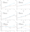
The role of TGF-β1 in bladder cancer cells was examined by observing cell viability by use of the MTT assay at 24, 48, and 72 hours after treating the bladder cancer cell lines 253J, 5637, T24, J82, HT1197, and HT1376 with TGF-β1. Among the six cell lines, 253J, 5637, HT1197, and HT1376 showed marked growth inhibition after TGF-β1 treatment. Although the T24 and J82 cell lines also showed inhibited cellular growth, the growth inhibition was less than that observed in the other four cell lines (Fig. 1). TGF-β1 did not stimulate proliferation of the cells but acted as a growth inhibitory factor in the bladder cancer cells. Overall, the TGF-β1 grossly inhibited cell growth in the bladder cancer cell lines. The pattern of inhibition depended on the type of cell line and the conditions used.
To double-check the growth inhibitory effect of TGF-β1 on bladder cancer cells, we examined the growth properties of 253J and T24 cells after neutralizing the action of TGF-β1 by using pan anti-TGF-β antibody. After pan anti-TGF-β antibody was added to the culture media, the growth properties of 253J and T24 cells that were suppressed by TGF-β1 were restored (Fig. 2). The regain of growth was remarkable at 72 hours in 253J cells and at 24 and 48 hours after TGF-β1 treatment in T24 cells (Fig. 2).
The 253J cells only and 253J cells with 4 ng/mL TGF-β1 were cultured in 1% serum media. The cells were harvested at 24, 48, and 72 hours to analyze the proportion of cells in a specific cell cycle and the proportion of cells experiencing apoptotic cell death. There were no significant differences in the proportion of cells in a specific cell cycle between the two treatments, with or without TGF-β1. However, there were more apoptotic cells in the TGF-β1-treated 253J cells (Fig. 3).
There are currently five distinct isoforms of TGF-β with 64% to 82% identity; only the TGF-β1, TGF-β2, and TGF-β3 forms are expressed in mammalian tissues [10]. Among these three isoforms, TGF-β1 is known as the predominant isoform in bladder tissues and cells in terms of protein and mRNA levels [11]. Thus, we chose TGF-β1 for the current experiment to elucidate the role of TGF-β in bladder cancer cells.
The role of TGF-β1 in bladder cancer cells was examined by observing cell viability by use of the MTT assay at 24, 48, and 72 hours after treating the 253J, 5637, T24, J82, HT1197, and HT1376 bladder cancer cell lines with TGF-β1. The TGF-β1 concentration used in this study (2 ng/mL and 4 ng/mL) was chosen on the basis of prior published studies [12,13] and a preliminary study performed in our laboratory (data not shown). For the cell viability study using the MTT assay, the 253J, 5637, HT1197, and HT1376 cell lines demonstrated inhibited growth after TGF-β1 treatment. The pattern of growth inhibition was time-dependent rather than dose-dependent. This pattern of dose independency has been observed in other cellular response experiments using growth factors [14,15]. These time-dependent inhibition patterns were clear at 48 hours after treatment (Fig. 1). Another two cell lines, T24 and J82, also showed inhibited cell growth, but the effect on growth inhibition was less than that observed in the 253J and 5637 cells (Fig. 1). A number of experimental studies have provided evidence for the acquisition of TGF-β1 resistance at relatively late stages of carcinogenesis and evidence that TGF-β1-resistant cancer often contains bioactive TGF-β1 [7]. It is reported that over-expression of TGF-β1 is associated with aggressive pathologic features, biological progression of disease, and death from bladder cancer [16]. We guessed that the cell lines established from invasive bladder cancer showed a proliferative response to TGF-β1 treatment before exhibiting the growth inhibition response of all bladder cancer cell lines. Among the cell lines used this work, all 5 cell lines except 253J originated from invasive bladder cancer; the 253J cell line was established from lymph node metastasis specimens [17,18]. These cellular response data suggest that TGF-β1 may be a candidate molecule for target therapy of aggressive bladder cancer. However, we must keep in mind that these are the results from an in vitro cellular response experiment only. Additional translational research is needed to apply this work to bladder cancer patients.
Among the six cell lines studied, the 253J and T24 cell lines showed reproducible results in repeated MTT assays. Therefore, we chose these two cell lines for further experimentation to double-check the growth inhibitory effect of TGF-β1. We neutralized the TGF-β1 effect by using the pan anti-TGF-β antibody and then observed growth patterns. The 253J and T24 cell lines were coincubated with TGF-β1 and the pan anti-TGF-β antibody. The addition of anti-TGF-β antibodies to the culture media restored the growth properties that had been inhibited by TGF-β1 (Fig. 2). Hence, these results provide evidence that the growth inhibition of bladder cancer cells was induced by TGF-β1.
Additional experiments were performed to study the mechanisms involved in growth inhibition. The 253J cell line was chosen because it showed constant and marked growth inhibition on repeat cell viability assays. In the FACS analysis, there were no significant differences in the cell cycle between the two treatments (the 253J cells only and the 253J cells with 4 ng/mL TGF-β1). However, there were more apoptotic cells in the TGF-β1-treated 253J cells. Therefore, TGF-β1 achieved growth inhibition by enhancing the level of apoptosis in the 253J cell line. It is known that TGF-β1 inhibits the growth of nonneoplastic epithelial cells by regulating molecules related to the G1 and S phases of the cell cycle. The cell cycle inhibition occurs through up-regulation of mito-inhibitors including p15, p21, and p27, and the cell cycle activation occurs through down-regulation of mito-activators including cyclins and cyclin dependent kinases [19,20,21,22]. Even though there are insufficient data on neoplastic cells, the mechanism for cell cycle regulation might be similar. In the present study, TGF-β1 induced growth inhibition of 253J cells and there were no significant differences in the cellular proportion of cell cycles (Figs. 1, 3). Even though TGF-β1 has been known as a micro-environmental regulatory molecule that signals cell cycle arrest, that feature was not evident in the bladder cancer cells. Whether there is a change in expression of the cell cycle regulation molecules after TGF-β1 treatment in bladder cancer cells would be valuable to study. The results of the present study seem to suggest that there might be no significant changes in the expression of the cell cycle regulation molecules in bladder cancer cells after TGF-β1 treatment. Recently, Al-Azayzih et al. [23] reported that TGF-β1 induces apoptosis via p38 mitogen-activated protein kinase and c-Jun N-terminal kinase/stress-activated kinase-mediated activation of caspases in T24 cells. This report strongly supports our opinion that TGF-β1 achieved growth inhibition by enhancing the level of apoptosis.
TGF-β1 did not stimulate cell proliferation but rather growth inhibition of bladder cancer cells. However, the pattern depended on the cell lines used. TGF-β1 achieved growth inhibition by enhancing the level of apoptosis in 253J cells. Overall, our data indicate that TGF-β1 can be considered as a candidate molecule for target therapy of bladder cancer.
Figures and Tables
FIG. 1
The cellular responses after transforming growth factor-β1 (TGF-β1) treatment in various bladder cancer cell lines. The cellular responses were examined at 24, 48, and 72 hours after treating the bladder cancer cell lines (T24, 253J, 5637, J82, HT1197, and HT1376) with TGF-β1. (A-F) All bladder cancer cell lines showed growth inhibition after TGF-β1 treatment. The properties of growth inhibition were more remarkable with the 253J, 5637, HT1197, and HT1376 cell lines (A, B, E, F) than the T24 and J82 cell lines (C, D) after the TGF-β1 treatment.

FIG. 2
The cellular responses after neutralizing with pan antitransforming growth factor-β1 (TGF-β1) antibody in the 253J and T24 bladder cancer cell lines. (A, B) After neutralizing the action of TGF-β1, the 253J and T24 cell lines recovered their proliferative function. In 253J cells, the recovery was remarkable 72 hours after the TGF-β1 treatment. (A) In T24 cells, the recovery was remarkable 24 and 48 hours after the TGF-β1 treatment (B).

FIG. 3
Fluorescence-activated cell sorter after the transforming growth factor-β1 (TGF-β1) treatment. There were more apoptotic cells in the TGF-β1 treated 253J cells. These findings were significant 48 hours after the TGF-β1 treatment (A). However, there were no significant differences in the cell cycle between the two treatments (B).

References
1. Blobe GC, Schiemann WP, Lodish HF. Role of transforming growth factor beta in human disease. N Engl J Med. 2000; 342:1350–1358.
2. Govinden R, Bhoola KD. Genealogy, expression, and cellular function of transforming growth factor-beta. Pharmacol Ther. 2003; 98:257–265.
3. Sanchez-Capelo A. Dual role for TGF-beta1 in apoptosis. Cytokine Growth Factor Rev. 2005; 16:15–34.
4. Roberts AB, Sporn MB. Transforming growth factors. Cancer Surv. 1985; 4:683–705.
5. Roberts AB, Wakefield LM. The two faces of transforming growth factor beta in carcinogenesis. Proc Natl Acad Sci U S A. 2003; 100:8621–8623.
6. Pasche B. Role of transforming growth factor beta in cancer. J Cell Physiol. 2001; 186:153–168.
7. Reiss M. TGF-beta and cancer. Microbes Infect. 1999; 1:1327–1347.
8. Kirkali Z, Chan T, Manoharan M, Algaba F, Busch C, Cheng L, et al. Bladder cancer: epidemiology, staging and grading, and diagnosis. In : Soloway M, Carmack A, Khoury S, editors. Bladder tumors. Paris: Health Publication Ltd;2005. p. 13–64.
9. Ministry of Health and Welfare, Korea Central Cancer Registry, National Cancer Center. Annual report of cancer statistics in Korea in 2010. Goyang: Ministry of Health and Welfare, Korea Central Cancer Registry, National Cancer Center;2011.
10. Roberts AB. Molecular and cell biology of TGF-beta. Miner Electrolyte Metab. 1998; 24:111–119.
11. Eder IE, Stenzl A, Hobisch A, Cronauer MV, Bartsch G, Klocker H. Expression of transforming growth factors beta-1, beta 2 and beta 3 in human bladder carcinomas. Br J Cancer. 1997; 75:1753–1760.
12. Park BJ, Park JI, Byun DS, Park JH, Chi SG. Mitogenic conversion of transforming growth factor-beta1 effect by oncogenic Ha-Ras-induced activation of the mitogen-activated protein kinase signaling pathway in human prostate cancer. Cancer Res. 2000; 60:3031–3038.
13. Champelovier P, El Atifi M, Mantel F, Rostaing B, Simon A, Berger F, et al. In vitro tumoral progression of human bladder carcinoma: role for TGFbeta. Eur Urol. 2005; 48:846–851.
14. Shin KY, Kim SJ, Choi WH, Choi DY, Park HY, Lee TY, et al. A study in the effects of epidermal growth factor and transforming growth factors-alpha on the growth of human transitional cell carcinoma cell lines. Korean J Urol. 1998; 39:45–50.
15. Iles RK. Ectopic hCGbeta expression by epithelial cancer: malignant behaviour, metastasis and inhibition of tumor cell apoptosis. Mol Cell Endocrinol. 2007; 260-262:264–270.
16. Kim JH, Shariat SF, Kim IY, Menesses-Diaz A, Tokunaga H, Wheeler TM, et al. Predictive value of expression of transforming growth factor-beta(1) and its receptors in transitional cell carcinoma of the urinary bladder. Cancer. 2001; 92:1475–1483.
17. Sanchez-Carbayo M, Socci ND, Charytonowicz E, Lu M, Prystowsky M, Childs G, et al. Molecular profiling of bladder cancer using cDNA microarrays: defining histogenesis and biological phenotypes. Cancer Res. 2002; 62:6973–6980.
18. Elliott AY, Cleveland P, Cervenka J, Castro AE, Stein N, Hakala TR, et al. Characterization of a cell line from human transitional cell cancer of the urinary tract. J Natl Cancer Inst. 1974; 53:1341–1349.
19. Ko TC, Sheng HM, Reisman D, Thompson EA, Beauchamp RD. Transforming growth factor-beta 1 inhibits cyclin D1 expression in intestinal epithelial cells. Oncogene. 1995; 10:177–184.
20. Geng Y, Weinberg RA. Transforming growth factor beta effects on expression of G1 cyclins and cyclin-dependent protein kinases. Proc Natl Acad Sci U S A. 1993; 90:10315–10319.
21. Ravitz MJ, Wenner CE. Cyclin-dependent kinase regulation during G1 phase and cell cycle regulation by TGF-beta. Adv Cancer Res. 1997; 71:165–207.
22. Carneiro C, Alvarez CV, Zalvide J, Vidal A, Dominguez F. TGF-beta1 actions on FRTL-5 cells provide a model for the physiological regulation of thyroid growth. Oncogene. 1998; 16:1455–1465.
23. Al-Azayzih A, Gao F, Goc A, Somanath PR. TGFβ1 induces apoptosis in invasive prostate cancer and bladder cancer cells via Akt-independent, p38 MAPK and JNK/SAPK-mediated activation of caspases. Biochem Biophys Res Commun. 2012; 427:165–170.


 PDF
PDF ePub
ePub Citation
 Print
Print


 XML Download