

 PDF
PDF ePub
ePub Citation
 Print
Print


INTRODUCTION
Prostate cancer (PCa) is the most common male neoplasm in North America. Since prostate-specific antigen (PSA) was introduced as a biomarker for PCa, the incidence of this disease has been increasing in many countries. However, PCa mortality decreased slightly in the mid to late 1990s in North America and several European countries [1]. Today, PCa tends to be diagnosed earlier and to be of lower grade than in past, and many patients prefer less invasive treatment modalities. One option is low-dose-rate brachytherapy (LDR-BT). This relatively noninvasive treatment modality involves the implantation of a low-energy radiation source within the prostate and has been used in the treatment of localized PCa for more than 3 decades [2]. In the past, ideal candidates for LDR-BT were considered to be subjects with low-risk PCa, that is, those with PSA<10 ng/mL, biopsy Gleason score (GS) ≤ 6, and clinical stage of T2a or less [3]. Indeed, there is no benefit of adding neoadjuvant or adjuvant hormonal therapy (HT) to LDR-BT in low-risk patients [4]. However, the indications for LDR-BT have recently been expanded to include intermediate- to high-risk cancers when administered in combination with some form of HT or external beam radiotherapy (EBRT) or both. The European Urological Association guideline described a possibility of LDR-BT in combination with supplemental EBRT or HT for intermediate- to high-risk PCa [5]. We can thus reasonably anticipate an increase in unexpected events relatively specific to aggressive PCa. The purpose of this study was to discuss safety and possible risks in expanding the indication of LDR-RT for higher-risk PCa on the basis of our own observational data. We analyze unusual events and focus discussion especially on pulmonary metastasis after LDR-BT.
MATERIALS AND METHODS
1. Patients
A total of 616 patients who had undergone LDR-BT at Jikei University Hospital between October 2003 and April 2010 were retrospectively reviewed in this study. The median follow-up of these patients was 48 months (range, 0-101 months).
All patients had histopathologically diagnosed PCa on the basis of prostatic needle biopsy. The absence of distant metastases was confirmed by chest x-ray, abdominal computed tomography (CT) scan, and bone scintigraphy. The cancer was staged in accordance with the unified tumor node metastasis system [6]. All slides of prostatic biopsy specimens were stained with hematoxylin and eosin and were reviewed by a single pathologist (H.T.). Tumor grade was determined according to the Gleason grading system. To evaluate cancer activity after biochemical recurrence (BCR), we used PSA kinetics as described by PSA doubling time (PSADT), which was calculated as log × 2 divided by the slope of the log PSA line (the difference in the 2 log PSA valued divided by the time between readings in months) [7]. Patients provided written informed consent before treatment.
2. Treatment
Patients were treated by ultrasound-guided implantation by using the Mick applicator as previously described [8]. Iodine-125 was used as the radiation source. The radioactive intensity of the implanted seed was 11.0 or 13.1 MBq. The prescribed doses of LDR-BT were 145 Gy for monotherapy and 110 Gy in combination with EBRT.
Japanese regulations specify the maximum permitted number of seeds for use and the maximum intensity of radiation [8,9]. To comply with these requirements in patients with relatively large prostate glands (≥40 cm3), we administered neoadjuvant hormonal therapy (NHT) for 3 to 6 months before brachytherapy. A combination of 1-month or 3-month depot injection of LHRH agonist (leuprolide acetate 3.75 mg or 11.25 mg, or goserelin acetate 3.6 mg or 10.8 mg), antiandrogen (bicalutamide 80 mg/d), or both were used for NHT. For patients with a higher GS, more advanced cancer, or higher PSA, we offered combined treatment with short-term NHT, EBRT, or both. These combined-treatment options were implemented before LDR-BT. EBRT was delivered by three-dimensional conformal or intensity-modulated radiation therapy. The treatment volume included the clinical target volume (the prostate, seminal vesicles, and internal margin) plus a 10-mm margin (5-mm posterior margin). The dose was prescribed to the isodose line covering the clinically planned target volume. The total EBRT dose was 39.5 to 46 Gy (median, 44 Gy). Nine-month adjuvant HT was administered in some patients as part of our multi-institutional randomized controlled study for intermediate-risk PCa (SHIP0804) [8]. One-month or 3-month depot injection of LHRH agonist alone was used for this purpose.
All patients underwent a CT scan for postimplant dosimetry 1 month after LDR-BT. The V100 index was measured as the percentage of target volume covered by the prescription dose. The histogram volume provided the dose delivered to 90% of the prostate (D90). We calculated the biologically effective dose with the use of an α/β ratio of 2 Gy for the purpose of comparison in total radiation dose [10].
Posttreatment follow-up was quarterly for the first 2 years and semiannually thereafter with a PSA measurement and rectal examination at each visit. Follow-up biopsy was performed when clinically indicated or routinely at 36 months after treatment in patients enrolled in the SHIP0804 study. BCR was defined as the PSA nadir plus 2 ng/mL by using the Phoenix definition [11]. Multiple imaging studies including chest x-ray, CT scan (chest, abdomen), bone scintigraphy, and pelvic magnetic resonance imaging, were applied in those with BCR in the search for clinical recurrence.
RESULTS
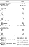
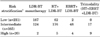
The patients' demographic data are shown in Table 1. The patients' median age was 69 years (range, 47-81 years) and their median initial PSA was 8.5 ng/mL (range, 2.1-34.6 ng/mL). The initial PSA was less than 10 ng/mL in 455 patients, 10 to 19.9 ng/mL in 149 patients, and at least 20 ng/mL in 12 patients. GS was less than 7 in 301 patients, 7 in 304 patients, and more than 7 in 11 patients. All patients had clinical stage T1 or T2 tumors. Risk stratification by the method of DAmico et al. [3] showed low-risk cancer in 231 patients, intermediate-risk cancer in 365 patients, and high-risk cancers in 20 patients. Table 2 shows the treatment according to the risk stratification. LDR-BT monotherapy was administered in 167 low-risk (72.3%), 124 intermediate-risk (34.0%), and 2 high-risk (10.0%) patients. Seventeen patients (4.7%) in the intermediate-risk group and 9 patients (45.0%) in the high-risk group received trimodality therapy with LDR-BT, HT, and EBRT. HT was performed 3 to 6 months before LDR-BT as NHT, and 43 patients (11.8%) in the intermediate-risk group received 9 months of additional HT after LDR-BT according to the SHIP0804 protocol [8]. Thus, among 616 patients, 269 were treated with HT and 80 were also treated with EBRT, including 26 treated with trimodality therapy. Average dosimetric parameters, with and without EBRT, were 95.9% and 95.3% for V100, 132.4 Gy and 164.4 Gy for D90, and 182.0 Gy2 and 173.8 Gy2 for BED, respectively.
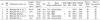
BCR occurred in 14 patients (6.1%) in the low-risk group, 25 patients (6.8%) in the intermediate-risk group, and 6 patients (30.0%) in the high-risk group during this follow-up period. Clinical recurrence, including the appearance of a solid mass at the site of a seminal vesicle or abdominal lymph node, bone metastasis, or pulmonary metastasis, was confirmed in 9 patients (64.3%) in the low-risk group, 13 patients (52.0%) in the intermediate-risk group, and 4 patients (66.7%) in the high-risk group. Five patients (19.2% of clinical recurrence) exhibited pulmonary metastases; 4 of these were isolated lesions with no involvement of any other sites (Table 3).
The individual demographics of the patients who developed pulmonary metastases are shown in Table 4. All of these patients had intermediate- or high-risk PCa. Median PSA was 8.2 ng/mL (range, 6.2-12.1 ng/mL), and clinical stage was T1c or T2a. GS was 7 or 8, including Gleason grade 4 in every patient. One patient received NHT and 3 received EBRT. Each received BED of 150 Gy2 or higher, ranging from 154.6 to 210.1 Gy2. The pulmonary metastases were diagnosed at a median follow-up of 24 months (range, 15-36 months). The PSADT before the appearance of pulmonary metastasis was short (median, 5.9 months; range, 2.7-9.7 months). All pulmonary masses subsequently showed regression after salvage HT; this confirmed the diagnosis (Fig. 1).
DISCUSSION
Pulmonary metastases from PCa are uncommon. Fabozzi et al. [12] reported radiological pulmonary metastases in 47 (3.6%) of 1290 PCa patients, but in most of these patients multiple distant metastases were also found in other locations, as in the end stage of the disease. Saitoh et al. [13] reviewed 1,885 autopsies of patients with PCa and found pulmonary metastasis in 49.1% of all metastatic disease. However, only 4 of 1,367 patients (0.3%) with distant metastases had isolated pulmonary lesions. Thus, these were extremely rare events [13]. Except for several case series, the world literature shows no systematic reviews of pulmonary metastases after definitive therapy for localized PCa [12].
Clinical recurrence was detected in 26 of 616 (4.2%) LDR-BT-treated patients in this study, involving seminal vesicles, abdominal lymph nodes, bones, and lungs. Five patients developed pulmonary metastases, including 4 cases (15.4%) of isolated lesions. The remaining patient had metastases at other sites also. This rate seems higher than the generally accepted low incidence of isolated pulmonary metastasis in localized PCa.
The etiology of pulmonary metastasis is difficult to ascertain but may result from the spreading of tumor cells into the bloodstream as the result of multiple puncture procedures during seed implantation. Transurethral resection of the prostate (TURP) has long been suggested as one of the causes of PCa dissemination [14]. Escape and migration of tumor cells through the rich venous plexus surrounding the prostate is one potential mechanism. Meacham et al. [15], however, monitored 379 patients treated with definitive radiotherapy for localized PCa that had been diagnosed by either needle biopsy or TURP and concluded that distant metastases after TURP may be due to poor prognosis in cases where tumors cause obstructive voiding symptoms rather than being a direct result of the resection.
Nevertheless, mechanical tumor dissemination is a possibility. For example, perineal needle tract seeding after 14G to 18G needle biopsy has been reported in 2% of PCa patients [16]. Seed migration to the lung during LDR-BT procedures has also been observed in 1.7% to 55.0% of patients [17,18]; placement of seeds near vascular structures may increase the probability of seed migration [17].
All these mechanisms may have contributed to tumor cell migration into the lung. We note that our data may be skewed; the patients who developed pulmonary metastases were among those we treated when we were still learning the techniques of brachytherapy (less than 30 months of experience). It is possible that our procedures were suboptimal, and that this facilitated tumor migration to the lung. Taussky et al. [19] reported the incidence of seed migration to decrease with accumulated experience. The overall incidence of seed migration was low, around 1%, in our series, and those with pulmonary metastases did not show evidence of seed migration.
It is also possible that preexisting pulmonary metastases were overlooked before LDR-BT, because only chest x-ray films were available at the time of clinical staging. However, we consider this a remote possibility, because the x-ray findings changed rapidly and remarkably over time after therapy.
Even if tumor migration occurs during LDR-BT, that migration itself cannot lead directly to metastasis. Multiple steps must occur, including the deposition of tumor cells in small vessels within the distant organ, extravasation into the surrounding tissue, and proliferation at the secondary site. At the same time, the tumor cells must successfully avert host immune responses and survive apoptotic signals [20]. All our patients with pulmonary metastasis had a high GS, reflecting biological aggressiveness with potentially more vital cancer stem cells [21]. Only one of our five patients with pulmonary metastasis underwent NHT before LDR-BT. Such therapy, and other more effective measures to prevent tumor dissemination during LDR-BT, may be of benefit for biologically aggressive tumors.
Although BCR, as evidenced by the magnitude of PSA change, indicates early recurrence of PCa, it is difficult to predict distant metastases based solely on PSA values. PSA kinetics, including the precise amount of change in PSA during a specific period after LDR-BT, has been reported to predict clinical recurrence. Forsythe et al. [22] noted the usefulness of PSADT for the detection of distant metastases following LDR-BT. They concluded that shorter PSADT and initial GS were predictors of distant metastasis. In our cases of pulmonary metastasis, 4 of the 5 patients showed PSADT of less than 6 months before the diagnosis of metastasis. A short PSADT after BCR may help to predict clinical recurrence, including pulmonary metastasis after LDR-BT. Admittedly, the distinction from PSA bounce phenomenon in such cases is a concern because distant metastases occurred at an early phase after LDR-BT in our study (median, 24 months).
Our findings are not compatible with more than three decades of world experience in modern LDR-BT. Lack of a control group and pulmonary biopsies is clearly a limitation, and our study may be criticized as being hypothesis-generating. Nevertheless, routine implementation of chest CT in the face of BCR in our study may have detected pulmonary metastases more efficiently. This possibility may have been overlooked owing to the liberal use of HT in such cases. This should be confirmed in a prospective series with larger numbers of patients.


 XML Download