

 PDF
PDF ePub
ePub Citation
 Print
Print


INTRODUCTION
Schwannomas are benign nerve sheath tumors composed of Schwann cells, which normally produce the insulating myelin sheath covering the peripheral nerves. Common locations include the head, neck, mediastinum, and retroperitoneum. Schwannomas are usually asymptomatic until they become large and compress the surrounding tissues. Most of them occur during the third and fourth decades of life, with an equal gender distribution [1]. We present the case of a schwannoma that originated in the scrotum.
CASE REPORT
A 45-year-old male presented with a history of gradually increasing, painless, left-sided testicular swelling lasting 6 months. On physical examination, a firm mass measuring 9.5 cm×9.5 cm was observed at the root of the scrotum, close to the testis. The patient had no neurological symptoms and careful evaluation for neurofibromatosis was negative. No lymphadenopathy was detected. The patient also had a friction ulcer over the scrotal skin and a provisional clinical diagnosis of an epididymal or testicular tumor was made.
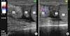
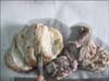
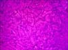
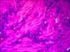
Ultrasonographic examination revealed a well-circumscribed heterogeneous scrotal mass that was well separated from the epididymis and testis and isoechoic in echotexture (Fig. 1). Preoperative evaluation for tumor markers of testicular tumors, which included alpha-fetoprotein, beta-hCG, and lactate dehydrogenase, were all negative. Curative surgical excision of the left scrotal mass was undertaken for removal of the tumor and a definitive diagnosis. Partial scrotectomy was done, which revealed a firm, nodular whitish lesion attached to the scrotal sac not involving the testis. The testis was spared and the scrotal mass was excised. The specimen submitted for histopathological examination consisted of an oblong, firm scrotal mass that measured 8.5 cm×8.5 cm×6.0 cm. The cut surface was whitish, lobulated, and glistening, with central yellow-brown areas (Fig. 2). Microscopic examination showed two different patterns: Antoni type A and Antoni type B areas. Antoni A areas are quite cellular and are composed of spindle cells that are often arranged in a palisading pattern or in an organoid pattern representing Verocay bodies. Antoni B areas are hypocellular with abundant edematous fluid-forming cystic spaces (Figs. 3, 4).
DISCUSSION
A schwannoma or neurilemmoma is a benign nerve sheath tumor composed of well-differentiated Schwann cells. The exact incidence of schwannomas is unknown, but they are rare. Although they are found in all age groups, schwannomas are more common in the first four decades of life and affect both sexes equally. More commonly, they are located in the head, neck, mediastinum, and retroperitoneum [2]. Rare sites reported include the face, neck, scalp, hands, tongue, palate, and larynx. Schwannomas may occur in association with neurofibromatosis or may arise sporadically. The scrotum is the rarest site for developing a schwannoma. To date, intrascrotal schwannomas have been reported only few times in the literature [1,3,4]. The microscopic appearance of a schwannoma is distinctive [5], with two recognizable patterns. Antoni A areas are composed of compacted spindle cells that are often arranged in palisades or in an organoid arrangement (Verocay bodies). Antoni B areas consist of tumor cells suspended in a myxomatous matrix that may appear microcystic.
Several variants of schwannomas based on appearances have been observed, including cellular, glandular, epithelioid, and ancient types, which exhibit benign progression. Cellular schwannomas are almost exclusively composed of Antoni A areas but lack Verocay bodies. The glandular and epithelioid variants are composed of epithelioid areas and glandular components, respectively, to acquire their descriptive names. Ancient schwannomas exhibit a bizarre hyperchromatic nucleus without mitosis [5]. Schwannomas pose a difficult diagnostic challenge to urologists. Radiological findings are often nonspecific [6]. Ultrasonography can differentiate between solid and cystic tumors. Computed tomography can be helpful in determining the size, location, local involvement, and distant spread. Magnetic resonance imaging provides similarly useful information to computed tomography, but yields better visualization of the tumor. Fine needle aspirate cytology is often not helpful because the tissue architectural information required cannot be obtained from cytological specimens [7]. The only gold standard diagnostic investigation is histology of either the biopsy or excised specimen. The mainstay of treatment remains surgical excision. Although malignant change is exceedingly rare [8,9], large and incompletely excised lesions are capable of recurrence. The present patient will require follow-up for a period of at least 2 years owing to the large size of the tumor and uncertainty of complete excision. Jiang et al. [10] in their study on male genital schwannoma followed the patients up for period for 2 years to 6 years (mean, 4.5 years). Extended follow-up beyond 2 years is required in cases having a large tumor size and in which the histopathology shows hemorrhage and necrosis.
 XML Download