

 PDF
PDF ePub
ePub Citation
 Print
Print


INTRODUCTION
Since Crocker first described extramammary Paget disease (EMPD) of the scrotum and penis [1], EMPD has been reported as a rare malignancy. Patients generally present with EMPD between the ages of 50 and 80 years. The disease is seen most frequently in Caucasians and more commonly in women than in men [2]. The most common sites of EMPD include the female genitalia and perianal regions; the penoscrotal area has been less commonly reported, with most cases described in Asia. Penoscrotal EMPD usually presents grossly as a well-demarcated erythematous lesion and symptoms like pruritus may precede the appearance of clinically visible lesions. Because of its rare incidence (1/3.7 million males annually) [3], the pathophysiology, staging, prognosis, and treatment of EMPD have not been clarified. The present study reports the clinical characteristics of EMPD of the penis and scrotum and the outcome of wide local excision of EMPD.
MATERIALS AND METHODS
A single-institution retrospective review identified 44 male patients who had visited Seoul National University Hospital (Seoul, Korea) for EMPD of the external genitalia between January 2000 and December 2012. Of these 44 patients, the analysis was performed for 19 patients who had undergone wide local excision. Excluded patients had undergone radiotherapy and chemotherapy without surgical excision or had undergone surgical excision in other hospitals. The study was approved by the Institutional Review Board granting approval at the institution.
The mean age of the patients at diagnosis was 68 years (range, 57 to 82 years). Most patients underwent computed tomography of the abdomen/pelvis, chest X-ray, and tumor marker evaluation (carbohydrate antigen 19-9, carcinoembryonic antigen [CEA], prostate-specific antigen, and alpha-fetoprotein). Patients in whom gastrointestinal malignancy was suspected underwent esophagoduodenoscopy and colonoscopy. Wide local excision was performed with or without preoperative mapping biopsy (Fig. 1) and intraoperative frozen biopsy, except in one patient who underwent wide local excision with grossly uninvolved lateral margins of 1 cm. In patients who did not undergo preoperative mapping biopsy, the initial excision margin was decided as 2 cm away from the grossly demarcated margin. All excisions were carried deep to the subcutaneous fat. Prophylactic lymph node dissection was not performed, except in one patient who was already known to have locoregional lymph node invasion without distant metastasis. Reconstruction was performed by split-thickness skin graft, full-thickness skin graft, local scrotal flap, or by primary closure.
All participants were followed up for a mean duration of 22.5 months (range, 1 to 60 months). Routine surveillance biopsies were not performed in asymptomatic patients. However, if any new suspicious lesions were found on physical examination, further punch biopsies were considered, and most patients underwent computed tomography or ultrasonography to evaluate for recurrence or progression.
The Fisher exact test was used for statistical analyses of factors described by p-value. A difference was considered statistically significant for p-values of less than 0.05. All statistical analyses were performed by using commercially available software (IBM SPSS ver. 18.0, IBM Co., Armonk, NY, USA).
RESULTS
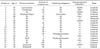
The clinical characteristics of the patients are shown in Table 1. All patients had presented with pruritus and erythematous lesions, which were the most common exam findings. In some patients, the EMPD lesions had been misdiagnosed as eczema or some other benign skin lesion at outside clinics and were thus managed with supportive treatment. This misdiagnosis delayed proper management by 43.5 months on average, ranging from 1 month to 198 months. To see whether this delay in treatment predicted worse outcomes, the patients were divided into two groups by a 30-month duration from onset of symptoms to treatment. This comparison did not reveal statistical differences in depth of invasion (p=0.108) or disease progression (p=0.263) between the early (<30 months) and delayed treatment (≥30 months) groups.
The lesions were located at the penoscrotal junction in 10 patients (52.6%), at the penoscrotal junction with spread to the pubic area in 4 (21.1%), at the scrotum in 4 (21.1%), and at the penis in 4 (4.8%). Of the 19 patients, 6 patients had presented to our institution with recurrent EMPD. Of these six, three patients had undergone the initial surgical excision at an outside institution. Among the remaining three patients, one had cryotherapy at an outside hospital, and two were treated with applications of imiquimod at the Department of Dermatology in our hospital. Of the whole, one patient had a locoregional invasive disease and another had metastatic disease at diagnosis, respectively. Three patients had underlying malignancies that were diagnosed and treated before the management of EMPD.
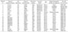
The details of treatment and outcomes in this cohort are summarized in Table 2. Except for one patient (No. 6), all of the patients had undergone preoperative mapping biopsy or intraoperative frozen biopsy (Fig. 1). Seven patients underwent preoperative mapping biopsy without intraoperative frozen biopsy. In seven patients, intraoperative frozen biopsy was positive and thus additional excisions were performed until a negative margin was obtained, except in one patient (No. 2) for whom margin-free resection would have created too large of a soft tissue defect. Tumor cells were detected in resected margins in 10 patients (52.6%) in final pathologic reports. Additional excision was not performed for these patients, who were closely followed up instead. There were four recurrent cases (21.1%), and two of these patients (10.5%) died from the disease. There were no recurrences in patients who underwent preoperative mapping biopsy. The performance of preoperative mapping biopsy was not correlated with the resection margin (p=0.628) or local recurrence (p=0.071). The lesion invaded the dermis in 14 patients (73.7%) and invaded the subcutaneous fat layer in 1 patient (5.3%). The resection margin (p=0.279) and invasion depth (p=0.235) did not correlate with local recurrence. In addition, invasion depth did not correlate with disease progression (p=0.532). Fig. 2 illustrates the cumulative incidence of recurrence and death.
Two of nine patients with a positive resection margin underwent additional excision of the positive lesion and there were no recurrences in these patients. Patients 2 and 3, in whom local recurrence was observed at 2 years and 8 months postoperatively, respectively, underwent secondary operations and have been followed up with no evidence of disease at 5 years. Patient 6 had undergone palliative chemotherapy with 5-fluorouracil and cisplatin but died from disease progression. Patient 18 had imiquimod treatment before the operation, but complete surgical excision of the large lesion could not be obtained owing to morbidity, including vessel injury, lymph edema, and large skin defect. His final pathologic report showed subcutaneous invasion. He underwent palliative radiation therapy but died of disease progression.
DISCUSSION
Mostly, EMPD arises as a primary cutaneous malignancy and rarely occurs in association with adnexal carcinoma of a nearby sweat gland or in association with a malignancy of an adjacent organ. Most reports of penoscrotal EMPD are from Asian cohorts [4-6]. Wide local excision has been thought to be the treatment of choice for penoscrotal EMPD. Yang et al. [5] reported that intraoperative frozen biopsy examination reduces rates of positive resection margins and recurrence. Our institute has performed intraoperative frozen biopsy or preoperative mapping biopsy since 2000. In the present study, we report the outcomes of penoscrotal EMPD patients who underwent surgical excision and review the clinical factors influencing outcomes.
The present study evaluated the efficacy of intraoperative frozen biopsy or preoperative mapping biopsy. No recurrence was shown in the latter group. Preoperative mapping biopsy can help surgeons to define excision margins for lesions and to decide on the reconstructive method. In contrast with previous studies [7] that reported that frozen biopsy improves the chance of obtaining negative margins, the resection margin was positive in 10 of 19 patients in the present study. Algaba et al. [8] reported that frozen biopsy could not guarantee negative resection margins. This may be because EMPD has multicentric origins with satellite lesions besides the gross lesion [4,9,10]. A positive resection margin was reported to be a risk factor for local recurrence [11], and depth of invasion and lymphovascular involvement were reported to be important pathological predictors of metastatic potential [7,12,13]. However, our findings did not show statistical significance, possibly because of the small sample size, and lymphovascular involvement was not included in the pathologic report, although two progressive cases had occurred among the 14 dermis-invasive diseases. The high proportion of dermis-invasive cases (14/19, 73.7%) is a salient point. This may reflect the health care trend in South Korea, by which most difficult cases are managed at tertiary university hospitals. In contrast with invasive disease, intraepidermal EMPD had a good prognosis in the present study, and no disease progression was observed despite two cases of local recurrence. Therefore, because EMPD is frequently mistaken for eczema or contact dermatitis, a biopsy should be performed on any uncontrolled inflammatory lesion of the external genitalia to prevent delayed diagnosis.
The pathogenesis of EMPD has not been elucidated, but the discussions of this have involved tumor cells spreading from apocrine or eccrine gland tumors, epidermotropic spreading from a regional malignancy, malignant transformation of epithelial cells, and malignant transformation of stem cells in the epidermis [5,6]. Internal malignancy was found to be a poor prognostic factor in a study of EMPD cohorts [3] in which 3 cases of internal malignancy were observed to have no local recurrence at follow-up. The incidence of internal malignancy related to EMPD is lower in Asians than in Caucasians [5,14]. Thus, routine colonoscopy to screen for EMPD-associated colon cancer may not be indicated among Asian patients. Instead, routine immunoperoxidase study for CEA and low-molecular-weight cytokeratin (CK) could be recommended if internal malignancy is suspected [2]. Most cases of EMPD revealed strong immunoreactivity for CEA and CK-7 with androgen receptor, in contrast with negative reactivity for CK-20 and p53. For instance, CK-20-positive EMPD may represent secondary Paget disease due to associated colorectal cancer, and C-erbB-2 was found to have a strong association with dermal invasion [15].
In the present study, cases treated by nonsurgical methods were not included. In the present study, a positive resection margin and local recurrence in intraepidermal cases had a relatively good prognosis. Among dermis-invasive cases, however, two patients with recurrence died of disease progression. In intraepidermal cases with local recurrence, wide excision of the lesion appears to be the most effective modality of treatment, as Li et al. [16] had reported. However, dermis-invasive lesions with local recurrence should be managed by wide excision with adjuvant management such as radiation therapy. Kim et al. [17] reported that radiotherapy can be effective for local control in unresectable cases, and Hata et al. [18] reported that radiotherapy is effective for curative treatment of EMPD. A retrospective review of the surgical management of EMPD from the Mayo Clinic [19] showed that Mohs surgery appears to be effective for Paget disease because the tumor has indistinct margins. Lee et al. [20] reported a high success rate of Mohs surgery for nonmetastatic EMPD in Korea. There are some studies on chemotherapy in the management of EMPD [21-25], but the efficacy has not been clarified. We plan to further investigate and report on the multimodality treatment of EMPD in a future publication.
CONCLUSIONS
Although penoscrotal EMPD is a rare malignancy, its diagnosis should not be delayed to allow for prompt treatment. Wide local excision is recommended with preoperative mapping biopsy and intraoperative frozen biopsy in nonmetastatic disease. Our findings suggest that intraoperative frozen biopsy or preoperative mapping biopsy cannot guarantee negative margins on final pathology. However, preoperative mapping biopsy and wide local excision with intraoperative frozen biopsy results in good prognosis in the treatment of EMPD, especially in those cases without dermal invasion. In recurrent cases, wide local excision is the treatment of choice, but adjuvant management like radiotherapy should be recommended in the presence of dermal invasion or regional invasion.


 XML Download