

 PDF
PDF ePub
ePub Citation
 Print
Print


INTRODUCTION
Elective nephron-sparing surgery for small renal masses (tumor size<4 cm, T1a) has been accepted as an oncologically safe alternative with limited complications in the presence of a healthy contralateral kidney [1]. The benefit of nephron-sparing surgery for small renal masses includes the preservation of renal function, with equal or better survival compared to radical nephrectomy [1,2]. Recently, the incidence of renal tumors including renal cell carcinoma (RCC) has been increasing around the world, which can be accounted for by increasing exposure to risk factors and increasing diagnosis of incidental tumors by use of improved imaging technology [3]. Parallel to the increase in incidentally discovered renal tumors, the size of tumors has become smaller [4,5].
Renal tumor size is important for the selection of a treatment modality and the prediction of prognosis. Previous studies have shown that the prognosis of RCC is dependent on the pathologic size of the tumor, especially for patients with tumors confined to the kidney [6,7]. However, treatment decisions, including the feasibility of nephron-sparing surgery, can only be made on the basis of the radiologic size of the tumor. Consequently, it is important to define the relationship between radiologic and pathologic size of renal tumors. Previous studies that examined the size difference between radiology and pathology yielded conflicting results [8-16]. In many of these studies, smaller tumor size and clear cell pathology were predictive of overestimated tumor size by radiology. To the best of our knowledge, studies comparing radiologic and pathologic tumor sizes according to tumor location are limited, and we could find only one report [16]. Therefore, in the present study, we examined the effects of different tumor conditions including tumor location (endophytic or exophytic) on discrepancy between the radiologically measured size and the true size of renal tumors to evaluate the appropriateness of the radiologically measured size in defining the criteria for nephron-sparing surgery.
MATERIALS AND METHODS
We retrospectively identified 217 consecutive patients who underwent radical or partial nephrectomy for a renal tumor suspected to be malignant at Ajou University Hospital between October 2003 and February 2011. None of the patients were diagnosed with von Hippel-Lindau disease, and none had received arterial embolization, targeted therapy, or immunotherapy before nephrectomy. All patients underwent a contrast-enhanced computed tomographic (CT) scan before surgery. The three-phase renal helical CT protocol used in our institution consists of an initial unenhanced scanning, followed by a corticomedullary phase, a nephrographic phase, and an excretory phase with 5-mm collimation. In case of a nonhelical CT scan taken at the referring hospital, the CT scan was repeated at our institution. The two CT scanners that are currently used in our hospital for the renal helical protocol are the Somatom Sensation 16-channel scanner (Siemens AG, Medical Solutions, Forchheim, Germany) and the Brilliance 64-channel scanner (Philips Medical Systems, Best, the Netherlands). Genesis Zeus (GE Healthcare, Milwaukee, WI, USA), which is a one-channel CT scanner using 7-mm collimation, was used interchangeably until 2005. We identified 36 cases evaluated with the Genesis Zeus scanner. We also identified 10 cases with only a CT scan taken outside our hospital. After the exclusion of these 46 patients, the remaining 171 patients were included in this study.
Radiographically, the tumor diameter was measured at various axes on a contrast-enhanced CT scan by one (K.B.L.) of the authors, and the largest of the diameters was taken to represent the radiologic size of the tumor. The pathologic tumor size was measured on gross surgical specimens before formalin fixation by a urologic pathologist. The surgical specimen was bisected along the longitudinal axis of the kidney, taking care to cut the tumor at the longest diameter. In case of an erroneous cut for an endophytic tumor, additional slices of the tumor were made to get a section at the largest diameter. The pathologic tumor size was taken as the largest diameter of the tumor measured on the bisected specimen.
The patients' demographic data were collected from the medical records. The histologic subtypes of tumor were categorized as clear cell RCC, papillary RCC, chromophobe RCC, unclassified RCC, oncocytoma, angiomyolipoma, and other benign tumors.
The preoperative CT scan was reviewed to categorize the tumor location. The tumor was arbitrarily classified as exophytic if more than 50% of the mass extended beyond the natural surface of the kidney and as endophytic if less than 50% of the mass extended beyond the natural surface of the kidney.
The mean values of the radiologic and pathologic size and the differences were calculated for the whole group and the different subgroups. If the sample size exceeded 30, normal distribution was assumed. In case of a sample size of 30 or smaller, the normality of the sample was tested by using the Shapiro-Wilk test. For normally distributed samples, the two measurements were compared by using the paired Student t-test. Otherwise, the two measurements were compared by using the Wilcoxon signed-rank test. SPSS ver. 16.0 (SPSS Inc., Chicago, IL, USA) was used for all statistics. Values of p<0.05 were considered to be statistically significant in all of the analyses.
RESULTS
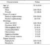
The mean age of the total 171 patients included in this study was 55.1±12.9 years. The patients included 120 men (70.2%) and 51 women (29.8%). Of the 171 tumors, 160 (93.6%) were RCC and 11 (6.4%) were benign tumors. Demographic data as well as pathologic data including the histologic subtypes of RCC and tumor location are shown in Table 1. Forty-four patients and 127 patients were evaluated with the 64-channel and the 16-channel CT scanner, respectively.
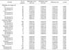
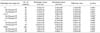
The mean radiologic and pathologic size for all tumors was 4.91±2.81 cm and 4.76±2.83 cm, respectively, and the difference was statistically significant (p=0.019) (Table 2). A comparison of the radiologic and pathologic tumor size in smaller subgroups of various categories is shown in Table 2. When subdivided into radiologic sizes of less than 4 cm, 4 to 7 cm, and larger than 7 cm, there was no statistically significant difference between the mean radiologic and pathologic sizes for tumors 4 to 7 cm (p=0.219) and larger than 7 cm (p=0.555). For tumors less than 4 cm, however, mean radiologic size was significantly larger than mean pathologic size (p=0.003). When subdivided according to histologic subtype, mean radiologic tumor size was significantly larger than mean pathologic tumor size only in clear cell RCC (p=0.002). When subdivided according to tumor location, mean radiologic tumor size was significantly larger than mean pathologic tumor size in endophytic tumors (p=0.043) but not in exophytic tumors (p=0.241). When endophytic tumors were categorized into radiologic sizes of less than 4 cm, 4 to 7 cm, and larger than 7 cm, there was a statistically significant difference between the mean radiologic and pathologic sizes for tumors less than 4 cm (p=0.001), but not for tumors 4 to 7 cm (p=0.073) and larger than 7 cm (p=0.603) (Table 3).
For all comparisons, a subanalysis was performed separately for each CT scanner. In all tumors, tumors less than 4 cm, clear cell RCC, and endophytic tumors, mean radiologic tumor size was significantly larger than mean pathologic tumor size, but the difference was statistically significant only for the 16-channel CT scanner and not for the 64-channel CT scanner (Tables 2, 3).
DISCUSSION
The increasing popularity of nephron-sparing surgery and other forms of ablative therapy for small renal masses has led to the necessity of creating valid tumor size criteria for selecting appropriate patients. In this respect, the adequacy of the 1997 TNM staging system has been challenged by many, and evidence has been presented that a tumor size cutoff of 4 or 4.5 cm has better prognostic value after radical nephrectomy than a tumor size cutoff of 7 cm [17,18]. Moreover, in a large series of 798 patients, Crispen et al. [19] showed that each 1-cm increment in tumor size from below 1 to 7 cm was associated with decreased long-term outcomes following partial nephrectomy, thus stressing the importance of tumor size in comparing outcomes following ablative and observational therapy. However, all these results were based on survival analysis of patients stratified by pathologic tumor size and not radiologic tumor size, when the latter is the actual guide to the treatment selection. For this reason, it is important to define the relationship between radiologic and pathologic sizes for localized renal tumors.
Some studies have examined the relationship between radiologic and pathologic sizes of renal tumors, and the results are conflicting. In 50 patients undergoing partial nephrectomy, Herr [8] prospectively compared the tumor size as assessed on a CT scan with the actual tumor size and showed that radiologic tumor size was 0.63 cm larger than pathologic tumor size. He concluded that, because renal artery occlusion results in shrinkage of the entire kidney, a similar decrease in tumor size might occur. From his observation, he suggested that partial nephrectomy could be attempted more often in patients with borderline tumor sizes. Irani et al. [9] retrospectively reviewed 100 patients with renal tumors who had undergone radical nephrectomy and also found that the average radiologic tumor size was significantly larger than pathologic tumor size (7 cm vs. 6 cm, p=0.005). Yaycioglu et al. [10] retrospectively reviewed 291 patients who underwent radical or partial nephrectomy and found that the difference between radiologic and pathologic size was not significant (p=0.1679). However, estimated blood loss of less than 700 mL, localized tumors, and clear cell RCC were associated with significantly larger radiologic size than the pathologic size. Interestingly, tumors that were more invasive and involved perinephric tissues had smaller radiologic size than pathologic size. According to the authors, this was caused by the close relation and mass effect of the adjacent structures in the more invasive tumors. In this context, larger estimated blood loss would be an indirect indicator of the invasiveness because of the more challenging dissection. They also warned that many additional features may lead to imprecise radiologic tumor measurement. Schlomer et al. [11] retrospectively identified 126 patients who underwent radical or partial nephrectomy within 60 days of CT scanning and found that the radiologic and pathologic sizes for all tumors were not significantly different (4.5 cm vs. 4.1 cm, p=0.35). However, in pathologic T1a tumors, the radiologic size was significantly larger than the pathologic size (p=0.009), and the difference was significant for tumors 4 to 5 cm in size (p=0.025). Kanofsky et al. [12] retrospectively studied 236 renal cancers and found that a reduction in tumor size owing to a loss of blood flow to the tumor had an impact on discordant radiologic and pathologic tumor sizes. In that study, the most frequent downstaging was observed for clear cell RCC compared with chromophobe or papillary type. Choi et al. [13] also found a large difference of greater than 0.45 cm between radiologic and pathologic renal tumor sizes, but only in pT1a, pT1b, and clear cell RCC and not in pT2 tumors.
In a more recent study, Mistry et al. [14] showed that there was no statistically significant difference between radiologic and pathologic sizes of renal tumor. In a retrospective analysis of 521 patients, Kurta et al. [15] found a statistically significant difference between the radiologic size (4.79 cm) and the pathologic size (4.69 cm) (p=0.02). However, the overall difference was only 1 mm, which suggests that CT scanning provides an accurate method of tumor size estimation. Lee et al. [16] retrospectively reviewed 467 patients who underwent radical or partial nephrectomy and found that the overall difference between radiologic and pathologic sizes was not significant except for tumors in the 4- to 5-cm range and for clear cell RCC. However, that difference was minimal and was judged to be clinically insignificant.
Our study showed a significant overall difference between radiologic and pathologic sizes (4.91 cm vs. 4.76 cm, p=0.019). When subclassified according to tumor size ranges and histologic subtypes, the difference was significant only for tumors less than 4 cm and for clear cell RCC. The reason for the significant size reduction only in the smaller tumor group is unclear. Central tumor necrosis is frequent in large tumors, which is often devoid of vasculature. This may lead to a lesser degree of tumor shrinkage following vascular occlusion in larger tumors. Frequent irregular tumor contours seen in larger tumors might make tumor size measurement more inconsistent. The small sample size of the larger tumors in our study might also explain the insignificant difference between the two measurements. Interestingly, the difference between the radiologic and pathologic sizes in our study became more significant when endophytic tumors were analyzed separately. This result is in contrast with the study of Lee et al. [16], which was the only study prior to ours to investigate radiologic and pathologic size differences by tumor location. Their study categorized tumors into exophytic, endophytic, and central (tumor completely buried within the renal parenchyma) and showed no significant tumor size difference according to the tumor location. Although the reason for the different results in the two studies remains uncertain, the different methods of categorization for the tumor location between the study by Lee et al. [16] and ours could have affected the results differently. We cannot disregard that, in a completely endophytic tumor, differentiation of the tumor margin from the surrounding parenchyma on a CT scan is more difficult and may lead to consistent overestimation. A completely endophytic tumor might also be under higher pressure from the surrounding parenchyma, resulting in a greater degree of tumor shrinkage following vascular occlusion. However, these hypotheses need to be validated by further studies.
For the sake of timeliness, we limited our study to tumors that were evaluated with contemporarily used CT scanners. To eliminate and observe potential intermethodological differences, we included CT scans taken only in our hospital and analyzed the results for the two different CT scanners separately. Although the 64-channel CT scanner showed insignificant differences between the radiologic and the pathologic tumor size, we believe that this result should be accounted for by the small sample size rather than by the superiority of the 64-channel CT scanner over the 16-channel CT scanner in terms of accuracy.
This study had several limitations. It was a retrospective, single-institution study, and the size of the study population was small. We did not analyze the effect of the two different measurement methods on patient prognosis. Although a significant size discrepancy existed between the two measurement methods in tumors less than 4 cm, we believe that the few millimeter difference in our relatively small sample size will not translate into a difference in prognosis. However, our results direct us to the need for a future study encompassing prognosis in a larger population with a longer follow-up. The tumor diameter measured in various axes on a CT scan may be inaccurate for the measurement of radiologic size in larger tumors owing to frequent irregular tumor contours.
Despite these limitations, we believe that a tendency exists to overestimate tumor size in smaller tumors and endophytic tumors on a CT scan. This may result in patients with a tumor size close to 4 cm undergoing radical nephrectomy instead of the more preferred nephron-sparing surgery. A better method of tumor size estimation will be necessary, especially for small tumors, which will result in better selection of patients who will benefit from nephron-sparing surgery.
CONCLUSIONS
Renal tumor size measured by contemporary CT scans seems to be overestimated in tumors less than 4 cm, in clear cell RCC, and in endophytic tumors. One reason the difference between mean radiologic and pathologic tumor sizes was statistically significant only for the 16-channel CT scan and not for the 64-channel CT scan may be the small sample size of the patients evaluated with the 64-channel CT scanner. Our results suggest that in planning a nephron-sparing surgery for renal tumors, especially for endophytic tumors of less than 4 cm, the size measured on a CT scan should be corrected to obtain a more precise estimate. A large-scale, multicenter, prospective study will be needed to confirm our findings and to define a better method of renal tumor size estimation.
 XML Download