

 PDF
PDF ePub
ePub Citation
 Print
Print


INTRODUCTION
Since the introduction of percutaneous nephrolithotomy (PNL) for the treatment of renal calculi by Fernstrom and Johansson [1], rapid progress has been made in the development of techniques for PNL [2]. Although some studies have reported better surgical outcomes for the management of renal calculi by use of shockwave lithotripsy (SWL) [3,4], PNL is still regarded as the standard treatment modality for management of renal calculi [5].
Various technical challenges have recently been introduced in relation to the methods of access and positioning of patients. Isac et al. [6] suggested that endoscopicguided renal access is safe and effective in comparison with the fluoroscopic-guided access technique. Sivalingam et al. [7] introduced a retrograde technique for establishment of a percutaneous tract for performance of PNL. DasGupta and Patel [8] reviewed various positions for performance of PNL, including the supine position.
According to these technological developments, many urologists have modified the standard methods of postoperative management, such as insertion of a nephrostomy tube. In a comparison of hospital stay and analgesic use, Nalbant et al. [9] demonstrated that totally tubeless PNL was a proper alternative to standard PNL. Considering the shorter hospital stay, Yun et al. [10] also emphasized that totally tubeless PNL was an effective alternative for the management of renal calculi. Using their experience of 3 years, Shah et al. [11] reported favorable outcomes of tubeless PNL.
However, the studies mentioned above have their limitations, and, to the best of our knowledge, no study of the feasibility of tubeless PNL for the management of renal staghorn calculi has been reported. Our study was designed to assess the feasibility of tubeless PNL by comparison of preoperative and postoperative parameters between conventional and tubeless PNL.
MATERIALS AND METHODS
Approval of the Institutional Review Board of Gachon University Medical Center (Incheon, Korea) was obtained before conduct of this study. A data set of all consecutive patients who underwent conventional or tubeless PNL for the treatment of renal staghorn calculi from 2003 to 2012 in a single center was retrospectively collected. Staghorn calculi were classified as a renal pelvis stone with branching into the major calyx. Also, stone burden was calculated as the surface area according to European Association of Urology guidelines [12]. All operations were performed by a single surgeon (H.J.) with the patient under general anesthesia.
Conventional and tubeless PNL were performed in the manner that we introduced previously [3]. Under general anesthesia, the patient's position was the lithotomy position. After insertion of an open-ended ureteral stent (6 Fr) via a cystoscope, the patient's position was changed to the prone position. By use of a fluoroscopic-guided "eye of the needle" technique [13], the affected kidney was punctured and an access tract was formed by using balloon dilation. Stone fragmentation was performed by using a lithoclast. In cases of conventional PNL, after removal of the stone, we placed a 24-Fr nephrostomy catheter. In cases of tubeless PNL, we inserted two pieces of Cutanplast (Mascia Brunelli, Italy) via the nephrostomy tract by using fluoroscopy instead of placing a nephrostomy catheter. After insertion of sealants, we checked for the presence of urinary leakage by infusion of contrast media by using an open-ended ureteral catheter.
We divided the patients into two groups. Group 1 included patients who underwent conventional PNL performed by a single surgeon (H.J.) for the treatment of renal staghorn calculi. Group 2 included patients who underwent tubeless PNL for the management of renal staghorn calculi performed by the same surgeon during the same period.
We compared various preoperative and postoperative parameters between the two groups. The preoperative parameters included age, sex, body mass index, stone laterality, and stone burden, and the postoperative variables included occurrence of complications and interventions and duration of hospital stay. We defined a stone-free state as no visible stones on a computed tomography scan at 1 month postoperatively.
Analyses of parameters of the two groups were performed by using an independent Student t-test and chi-square test. A p-value <0.05 were considered as having statistical significance. All statistical analyses were performed by using SPSS ver. 12.0 (SPSS Inc., Chicago, IL, USA).
RESULTS
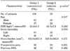
A total of 165 patients were enrolled in our study. Group 1 (n=106) included 61 male patients and 37 female patients. No significant differences in sex were observed between group 2 (n=59) and group 1. Body mass index (±standard deviation) was 24.0±3.5 and 24.7±3.2 kg/m2 in groups 1 and 2, respectively (p=0.24). In the analysis of demographic parameters, no significant differences in stone laterality or stone burden were observed between the two groups. Although no significant difference in stone burden was observed between the two groups, the presence of preoperative pain was significantly greater in group 1 than in group 2 (92 vs. 38, p=0.001). Twenty-six patients in group 1 and 19 patients in group 2 underwent SWL before PNL (Table 1).
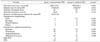
Table 2 shows various intraoperative and postoperative parameters of both groups. Mean operation times were 208.7±72.1 and 169.9±45.1 minutes in groups 1 and 2, respectively. In group 1, the mean duration of nephrostomy catheter indwelling was 3.55±1.94 days. Although no significant difference in the stone-free clearance rate was observed between the two groups, the mean hospital stay was significantly shorter in group 2 than in group 1 (5.32±2.37 vs. 7.08±6.34 days, p=0.01).
In the analysis of postoperative complications, fever episodes were more frequent in group 2 than in group 1 (13 vs. 5, p=0.01). However, almost all cases of fever in group 2 were due to atelectasis related to the general anesthesia and were easily managed with lung care. No significant differences in other complications were observed between the two groups.
For management of remnant stones, 26 and 13 patients in group 1 and group 2 underwent reoperation by PNL or SWL, respectively (p=0.849).
DISCUSSION
PNL was first introduced in 1976 by Fernstrom and Johansson [1] However, after several years, extracorporeal SWL was developed as a noninvasive technique for the treatment of renal and ureteral stones. Owing to its invasiveness by puncture of the renal parenchyme, postoperative complications after PNL have been reported [14,15]. Because of the noninvasiveness of SWL, many studies have supported the feasibility of SWL as an alternative to PNL [3,4]. Nonetheless, there is no doubt that PNL is still the standard method for the management of renal staghorn calculi [2,5].
In the process of performance of standard PNL, insertion of a nephrostomy catheter via a working track is inevitable for control of renal parenchymal bleeding and urinary leakage. Unfortunately, indwelling of the nephrostomy catheter is the primary reason for discomfort in patients postoperatively and prolongation of hospital stay.
According to advancements in the technique, many studies have reported on the feasibility of tubeless PNL [16,17]. Considering the length of hospital stay and analgesic use, Nalbant et al. [9] suggested that the tubeless PNL technique is more effective than conventional PNL. In addition, in a comparison of the data on conventional and tubeless PNL, Yun et al. [10] supported the same idea. However, the above studies had some limitations owing to small sample sizes and selection of patients.
Shah et al. [11] reported on their 3 years of experience with 454 cases of tubeless PNL. Despite their large samples sizes, their study also had some limitations because complicated cases, including those with pyonephrosis, intraoperative bleeding, and incomplete operations, were excluded from the tubeless PNL group.
In our study, the selection of all patients was performed consecutively. Group 1 included 106 patients who underwent conventional PNL from 2003. After the introduction of the tubeless PNL technique, another 59 patients underwent PNL using a tubeless method. All operations were performed by a single surgeon (H.J.). Accordingly, considering the single surgeon and the consecutive enrollment of patients, statistical bias was lower in our study.
Our results showed that tubeless PNL is a feasible and beneficial technique, considering the shorter duration of hospital stay and equal safety, compared with conventional PNL. The same number of complications in postoperative bleeding and wound infection were observed in groups 1 and 2. Although a higher incidence of fever was reported in group 2 than in group 1, in almost all cases, the febrile episodes were due to atelectasis related to the general anesthesia. However, differences in the operation time between the two groups were assumed to be due to the operator's learning curve according to the accumulation of cases rather than to the excellence of tubeless PNL.
Our study also had some limitations. First, our study was conducted retrospectively. However, all data were collected prospectively because the patients were enrolled in a consecutive manner. Second, the tubeless PNL group (group 2) did not include patients who underwent the procedure by use of a totally tubeless method, which refers to the use of no ureteral stents or nephrostomy catheters. In our study, totally tubeless PNL was applied to approximately two thirds of patients, whereas recent studies have reported results using totally tubeless PNL. Nonetheless, because there was no selection bias, our study has some significance.
Considering the weak and strong points of various studies related to tubeless PNL, including our study, the tubeless PNL technique may be a feasible and effective technique, compared with conventional PNL, in terms of shorter hospital stay and an equally proven complication rate. Our future study will focus on comparison of the results between tubeless PNL with ureteral stenting and totally tubeless PNL.
CONCLUSIONS
In the current study, our findings showed that tubeless PNL had the same safety and effectiveness as conventional PNL. According to our results, it is highly suggestive that tubeless PNL may be a feasible and safe alternative to conventional PNL for proper management of renal staghorn calculi.
 XML Download