Journal List > Korean J Urol > v.54(9) > 1006013

Seklehner, Fellner, Engelhardt, Schabauer, and Riedl: Percutaneous Radiofrequency Ablation of Renal Tumors: A Single-Center Experience

Abstract

Purpose

To evaluate the oncological outcomes, complications, and changes in renal function in patients treated with computed tomography-guided percutaneous radiofrequency ablation (RFA) for small renal tumors.

Materials and Methods

The charts of patients who underwent RFA from 2006 to 2011 at a single institution were reviewed. Oncological and functional outcomes were assessed. Statistical analyses were performed with IBM SPSS ver. 18.0 (IBM Co., Armonk, NY, USA).

Results

A total of 44 RFAs were done in 40 patients. Biopsy prior to RFA was performed in 79.6% of procedures. Of those, 68.6% had renal cell carcinoma (RCC). Mean tumor diameter was 26.2 mm. Grade I complications occurred in 25% of cases (n=11, pain or elevated temperature) and grade II complications in 2.3% (n=1, perirenal bleeding needing two units of blood transfusion). Serum creatinine slightly increased by 0.14 mg/dL at 2 years after RFA (p<0.004). Tumor recurrences were suspected in 8 of 43 cases during follow-up. In five patients, the suspected recurrence was a false-positive as shown by a negative biopsy result or lack of contrast enhancement on subsequent imaging. The verified recurrence rate was 7.7% in all tumors and 2.5% in RCC at a mean follow-up of 2 years. Tumor-free survival was 90% in all patients and 87.5% in those with RCC. Metastasis-free survival was 97.5% and cancer-specific survival was 100%.

Conclusions

Percutaneous computed tomography-guided RFA shows promising results at intermediate follow-up. Suspected tumor recurrences are frequently false-positives findings. A longer follow-up is required to verify the durability of these results.

INTRODUCTION

Renal cell carcinoma (RCC) is among the most frequent malignant tumors with significant morbidity and mortality. More than 58,000 estimated new cases and more than 13,000 deaths occurred in the United States in 2010 [1]. During the last decades, an increase in the incidence of all clinical stages of renal tumors was observed, with the greatest increase for localized tumors [2]. Owing to the wide use of cross-sectional abdominal studies such as ultrasound, computed tomography (CT), and magnetic resonance imaging (MRI), the detection rate of small solid lesions has increased, with up to 66% of tumors found incidentally [3]. The majority of incidentally diagnosed RCC tends to be of smaller size and thus is more likely to be asymptomatic, show a lower histological grade, and have a decreased incidence of metastasis [4].
Radiofrequency ablation (RFA) is a novel minimally invasive therapeutic approach that should be offered to patients with small renal tumors with a size less than 4 cm in diameter or significant comorbidities precluding surgical resection [5]. In the need for a therapeutic approach for such selected cases, RFA was established at our institution in 2006. In the present study we sought to assess the efficacy, complications, and changes in renal function in our initial cases after an intermediate follow-up period.

MATERIALS AND METHODS

We reviewed the charts of patients who underwent RFA between 2006 and 2011. Percutaneous RFA was offered to highly selected patients whose renal tumors did not exceed 40 mm in diameter. Patient selection was limited to subjects with advanced age and severe comorbidities that would cause a high surgical risk, impaired renal function prior to treatment, a functional or anatomical solitary kidney, or bilateral renal tumors or patients who refused tumor resection.

1. Renal biopsy and RFA procedure

After an initial implementation and learning process during which no renal biopsies were done, biopsies were routinely performed a few days before RFA under CT guidance. Biopsies were taken with an 18-Fr needle under local anesthesia. The specimens were fixed with hematoxylin-eosin staining.
All RFAs were performed under general anesthesia with a Rita device (Model 1500 RF Generator, 25 cm StarBurst XL Semi-Flex RFA Device, Angiodynamics, Queensbury, NY, USA) by an interventional radiologist. According to the kidney protocol of the Rita device, the maximum power to achieve a target temperature of 105℃ was 150 W. Depending on the target size, the time of each cycle varied. For a desired ablation defect of 20 mm, we used 5 minutes at the target temperature with a reset time of 5 minutes with a second identical cycle. For a 30-mm defect, we analogously used 7 minutes, and for a 40-mm defect, 8 minutes. If necessary, overlapping ablations were performed by repositioning the probe and restarting the procedure. At the end of the ablation, after the probe had been removed, a control CT scan verified the ablation and excluded complications.
An overnight stay at the hospital was mandatory for all patients. Patients were examined the day after the procedure through physical examination, ultrasound, and blood samples. The Clavien-Dindo classification was used to assess RFA-related complications [6].
The patients' renal function was assessed immediately before RFA, the day after RFA, and at follow-up by estimating the glomerular filtration rate (eGFR) with the MDRD equation, as modified in 2005: eGFR=175×(creatinine) -1.154×(age)-0.203×(0.742 if female).

2. Definitions of oncological outcome and follow-up schedule

Four definitions of treatment outcomes were used: complete and incomplete treatment and suspected and verified recurrence. Complete treatment was defined as a lack of contrast enhancement in combination with shrinkage or a stable size of the ablated tumor at the first follow-up MRI/CT. Conversely, incomplete treatment was defined as contrast enhancement or progression in tumor size at the first follow-up imaging study after treatment. Suspected tumor recurrence was defined as contrast enhancement or extension in size in any subsequent imaging in initially completely treated patients. Suspected tumor recurrence turned into a verified recurrence either when a renal biopsy of the lesion was positive for vital tumor tissue or when the site of the formerly ablated lesion further increased.
Patients with RCC or an unknown histology were followed with contrast MRI or CT every 3 months after treatment in the first year and then every 6 months, whereas oncocytoma patients were followed every 6 months.

3. Ethical concerns and statistical analysis

This retrospective study was conducted in accordance with good clinical practice guidelines. All patients signed an informed consent before treatment. The principles of the Helsinki Declaration were followed.
Statistical analyses, including the related-samples Wilcoxon signed-rank tests and the chi-square test, were performed with IBM SPSS ver. 18.0 (IBM Co., Armonk, NY, USA). A p-value of<0.05 was considered significant.

RESULTS

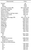
A total of 44 percutaneous RFA procedures were performed in 40 patients. Mean tumor size was 26.2 mm (range, 15 to 42 mm), mean length of hospital stay was 1.4 days (range, 1 to 4 days), and mean follow-up was 23.8 months (range, 3 to 59 months). Patient and tumor characteristics are provided in Table 1.
Thirty-five of the 44 tumors were biopsied before RFA (79.6%). In biopsied patients, RCC was the prevalent histology in 68.6% and oncocytoma was the prevalent histology in 14.3%; 17.1% of cases were benign or inconclusive.

1. Complications

Grade I complications occurred in 11 patients (25%) and grade II complications in 1 case (2.3%). All grade I complications (pain or elevated temperature) were treated conservatively with anti-inflammatory drugs, whereas the one patient with the grade II complication (perirenal bleeding) received two units of blood without the need for any further surgical interventions (Table 2).

2. Renal function

Overall changes in renal function and a precise breakdown of every patient are shown in Table 3. At more than 2 years after the treatment, the mean serum creatinine increased by 0.14 mg/dL on average (p<0.004). Stratified by length of follow-up, the increase was not significant in the first 24 months but was after 24 months (difference, 0.24 mg/dL; p<0.001). Mean eGFR decreased significantly by 7.5 mL/min from 65.9 mL/min before treatment to 58.4 mL/min at maximum follow-up (range, -55.2 to 17.3 mL/min; standard deviation [SD], 13.04; p=0.004). The distribution of patients in the corresponding eGFR subgroups (≥60, 59-30, 29-15, and ≤15 mL/min) did not change significantly from before therapy to maximal follow-up (p=0.08).

3. Complete and incomplete treatment

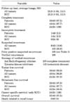
Forty-three of 44 ablated tumors (97.5%) were classified as completely treated on the first imaging study at follow-up, which normally took place 1 to 3 months after RFA (Table 4). One ablation was incomplete owing to anatomical limitations during RFA. This patient is currently under active surveillance with no sign of tumor progression or recurrence.

4. Suspected and real tumor recurrence, its management, and overall outcome

In 8 of 43 cases (18.6%) initially classified as complete ablations, radiologists suspected tumor recurrences during follow-up. The suspected recurrences were observed after an average of 23.9 months (range, 11 to 43 months) and had an average size of 22.6 mm (range, 15 to 30 mm; SD, 7.0). Of those eight suspected recurrences, five (62.5%) turned out to be false-positives whereas three (37.5%) were confirmed (Table 5).
The false-positive recurrences showed a negative (repeat) biopsy or, if the patient refused biopsy, showed no further contrast enhancement on subsequent imaging (10 or 12 months after suspected recurrence). Average follow-up until occurrence of the false-positive recurrences was 25.2 months (range, 11 to 43 months).
The three proven recurrences (7.7%) were noted at 12, 23, and 30 months after RFA. Two proven recurrences were treated with a second RFA with complete ablation. One patient developed metastatic disease and is currently being treated with tyrosine kinase inhibitors.
In our cohort, the overall survival was 87.5%; five patients died from comorbidities, and renal tumors were not responsible for their deaths. In patients with verified RCC, the tumor-free survival rate was 90% and the metastasis-free survival rate was 95.8% at a mean follow-up of 23.3 months.

DISCUSSION

Ablative techniques like percutaneous RFA are emerging because they offer an alternative to surgical excision, especially for elderly patients with impaired health. With the better understanding of the growth kinetics and behavior of small renal tumors [7-9], ablative procedures have become of particular interest.
Most small renal tumors have a slow growth rate and rarely metastasize [7]. Tumor size seems to be a predictor of the tumor growth rate [7], whereas size alone is an insufficient parameter to distinguish between RCC with a so-called benign biological behavior from one with an aggressive behavior [8]. Others have reported that the aggressive potential of RCC increases beyond a diameter of 30 mm [9].
Imaging is not presently able to assess the malignancy of renal tumors: only angiomyolipoma can be diagnosed with sufficient accuracy [10]. Renal biopsy is a safe procedure with an incidence of grade I complications of about 10% [11]. Biopsy success depends on tumor size and is diagnostic in 81% in tumors with an average size of 25 mm (showing RCC in about 75% and benign histology in about 20%) [11]. A repeat biopsy, after an initial nondiagnostic one, provides similar diagnostic rates so that diagnosis for most patients can be achieved [11].
In terms of avoiding unnecessary and unjustified procedures owing to benign, unknown, or inconclusive histology, we recommend our strategy of taking a renal biopsy under local anesthesia a few days before RFA. With a biopsy rate of nearly 80%, our study differs from most of the previously published studies [12-19].
Unlike most previously published series [12-22] (Table 6), we used a generally accepted grading system of complications, thus guaranteeing transparency and comparability [9]. The minimally invasive nature of this procedure was confirmed by the low rate of complications in our setting: only minor complications occurred in 27% (grade I, 25%; grade II, 2.3%). All complications were treated conservatively without the need for surgical intervention. RFA is associated with a significantly lower incidence of complications, morbidity, and mortality compared with tumor resection, especially laparoscopic nephron-sparing surgery [5]. Complications after partial or radical nephrectomy are more likely to occur in older patients with preexisting comorbidities, and postoperative complications after nephrectomy are associated with a significantly higher risk of death [23]. This should be taken into consideration when patients with comorbidities present with small asymptomatic renal tumors.
The increase of serum-creatinine by 0.14 mg/dL at a 2-year follow-up and a slight decrease in eGFR conforms to the literature [24,25]. Ablative techniques have little impact on renal function in subjects with regular renal function as well as in those with a solitary kidney or renal insufficiency [24]. Renal function is diminished less by ablative procedures than by partial or radical nephrectomy [15]. This is of particular interest because the association between impaired renal function and cardiovascular morbidity and mortality is well established [26].
Of note, we experienced a considerable discrepancy in suspected and proven recurrences during follow-up. Recurrences of initially complete ablations were suspected in eight patients. Interestingly, 62.5% of all suspected recurrences (mean follow-up, 25.2 months) showed no evidence of malignancy in the (repeat) biopsy or no further contrast enhancement in subsequent imaging under active surveillance. In one case, renal biopsy identified a focal inflammation that caused the contrast enhancement. The reason for the remaining four false-positive recurrences remains unknown. A possible explanation could be imaging artifacts or inconclusive biopsies. However, all images were reviewed by radiologists experienced in post-RFA imaging. It is known that renal biopsies are nondiagnostic in up to 19% of cases [11].
The considerable number of false-positive suspected recurrences must be emphasized, because immediate retreatment, either with a second ablation or even with tumor resection, might be an unnecessary overtreatment. In cases of suspected recurrences, a repeat biopsy or even temporarily active surveillance in selected patients might be justified, especially in small, asymptomatic tumors. Breda et al. [27] stated that active surveillance up to 1 year after post-therapeutic enhancement could be justified, because most of these enhancements are not a sign of recurrence but a result of postoperative inflammation immediately after RFA. However, their results relate to enhancements immediately after RFA and not to events occurring 11 to 43 months after RFA.
The present study had several limitations. First, it was retrospective and nonrandomized in nature. Second, not every patient underwent renal biopsy before RFA or if a recurrence was suspected. This was related to multiple factors such as the learning curve, the implementation process of RFA in our department, and patient's choice. However, this was mitigated because we separately analyzed patients undergoing RFA with biopsy-proven RCC. Third, renal function was assessed with serum creatinine and eGFR and not with more accurate measures such as diethylene triamine pentaacetic acid-scans, thus diminishing the validity of our results in this field. The primary intention of the study was to report the efficacy of RFA in highly selected patients who were offered a treatment option and not to assess the influence of RFA on renal function. Finally, follow-up was limited to a mean of 2 years; changes in tumor-free survival or renal function might occur later on.
Owing to demographic changes, the increased incidence of renal tumors in the elderly [28], a better understanding of the growth kinetics and behavior of small renal tumors [7-9], the low rate of complications [14,16-18,20-22], and the only slight impairment of renal function [15,26], the role and importance of RFA might further increase in the future.

CONCLUSIONS

Percutaneous CT-guided RFA shows promising results after intermediate follow-up. Suspected tumor recurrences are frequently false-positives. A longer follow-up is required to verify the durability of these results.

Figures and Tables

TABLE 1
Patient and tumor characteristics
kju-54-580-i001

Values are presented as number (%) unless otherwise indicated. SD, standard deviation; eGFR, estimated glomerular filtration rate.

TABLE 2
Complications related to percutaneous radiofrequency ablation by utilizing the Clavien-Dindo classification
kju-54-580-i002
TABLE 3
Renal function before and after RFA
kju-54-580-i003

Table 3 shows the mean renal function before and after RFA. The impact of RFA on renal function was minor. Serum creatinine just slightly decreased during follow-up (p<0.004) but the distribution of patients according to the eGFR classification remained stable (p=0.080).

RFA, radiofrequency ablation; eGFR, estimated glomerular filtration rate; SD, standard deviation.

TABLE 4
Outcome after percutaneous radiofrequency ablation
kju-54-580-i004

Values are presented as number (%).

Only one treatment was classified as incomplete treatment whereas 97.5% as complete treatment at first follow-up imaging. It is remarkable that eight tumors were suspected to be a recurrence at a later stage of follow-up. Of note, only three of those suspected recurrences were confirmed, leading to the shown rates of cancer specific, tumor free, metastasis free and overall survival. RCC, renal cell carcinoma; SD, standard deviation.

TABLE 5
False positive suspected recurrences
kju-54-580-i005

The table summarizes all patients, whose suspected tumor recurrences were not confirmed: three patients had a negative biopsy of the lesion and two patients, who refused biopsy, showed no further contrast enhancement at subsequent imaging under active surveillance.

RFA, radiofrequency ablation; CT, computed tomography; RCC, renal cell carcinoma; MRI, magnetic resonance imaging.

TABLE 6
Literature review of percutaneous RFA
kju-54-580-i006

The table pgrovides a literature review of published studies assessing complications and efficacy of percutaneous RFA.

RFA, radiofrequency ablation; RCC, renal cell carcinoma; pt, patient.

Notes

The authors have nothing to disclose.

References

1. Jemal A, Siegel R, Xu J, Ward E. Cancer statistics, 2010. CA Cancer J Clin. 2010; 60:277–300.
2. Hock LM, Lynch J, Balaji KC. Increasing incidence of all stages of kidney cancer in the last 2 decades in the United States: an analysis of surveillance, epidemiology and end results program data. J Urol. 2002; 167:57–60.
3. Volpe A, Panzarella T, Rendon RA, Haider MA, Kondylis FI, Jewett MA. The natural history of incidentally detected small renal masses. Cancer. 2004; 100:738–745.
4. Polascik TJ, Mouraviev V. The rise of ablative technologies for treating the small renal mass. Eur Urol. 2007; 52:636–638.
5. Ljungberg B, Cowan NC, Hanbury DC, Hora M, Kuczyk MA, Merseburger AS, et al. EAU guidelines on renal cell carcinoma: the 2010 update. Eur Urol. 2010; 58:398–406.
6. Dindo D, Demartines N, Clavien PA. Classification of surgical complications: a new proposal with evaluation in a cohort of 6336 patients and results of a survey. Ann Surg. 2004; 240:205–213.
7. Mason RJ, Abdolell M, Trottier G, Pringle C, Lawen JG, Bell DG, et al. Growth kinetics of renal masses: analysis of a prospective cohort of patients undergoing active surveillance. Eur Urol. 2011; 59:863–867.
8. Klatte T, Patard JJ, de Martino M, Bensalah K, Verhoest G, de la Taille A, et al. Tumor size does not predict risk of metastatic disease or prognosis of small renal cell carcinomas. J Urol. 2008; 179:1719–1726.
9. Remzi M, Ozsoy M, Klingler HC, Susani M, Waldert M, Seitz C, et al. Are small renal tumors harmless? Analysis of histopathological features according to tumors 4 cm or less in diameter. J Urol. 2006; 176:896–899.
10. Israel GM, Bosniak MA. Pitfalls in renal mass evaluation and how to avoid them. Radiographics. 2008; 28:1325–1338.
11. Leveridge MJ, Finelli A, Kachura JR, Evans A, Chung H, Shiff DA, et al. Outcomes of small renal mass needle core biopsy, nondiagnostic percutaneous biopsy, and the role of repeat biopsy. Eur Urol. 2011; 60:578–584.
12. Altunrende F, Autorino R, Hillyer S, Yang B, Laydner H, White MA, et al. Image guided percutaneous probe ablation for renal tumors in 65 solitary kidneys: functional and oncological outcomes. J Urol. 2011; 186:35–41.
13. Tracy CR, Raman JD, Donnally C, Trimmer CK, Cadeddu JA. Durable oncologic outcomes after radiofrequency ablation: experience from treating 243 small renal masses over 7.5 years. Cancer. 2010; 116:3135–3142.
14. Varkarakis IM, Allaf ME, Inagaki T, Bhayani SB, Chan DY, Su LM, et al. Percutaneous radio frequency ablation of renal masses: results at a 2-year mean followup. J Urol. 2005; 174:456–460.
15. Lucas SM, Stern JM, Adibi M, Zeltser IS, Cadeddu JA, Raj GV. Renal function outcomes in patients treated for renal masses smaller than 4 cm by ablative and extirpative techniques. J Urol. 2008; 179:75–79.
16. Levinson AW, Su LM, Agarwal D, Sroka M, Jarrett TW, Kavoussi LR, et al. Long-term oncological and overall outcomes of percutaneous radio frequency ablation in high risk surgical patients with a solitary small renal mass. J Urol. 2008; 180:499–504.
17. Turna B, Kaouk JH, Frota R, Stein RJ, Kamoi K, Gill IS, et al. Minimally invasive nephron sparing management for renal tumors in solitary kidneys. J Urol. 2009; 182:2150–2157.
18. Park S, Anderson JK, Matsumoto ED, Lotan Y, Josephs S, Cadeddu JA. Radiofrequency ablation of renal tumors: intermediate-term results. J Endourol. 2006; 20:569–573.
19. Kim JH, Kim TH, Kim SD, Lee KS, Sung GT. Radiofrequency ablation of renal tumors: our experience. Korean J Urol. 2011; 52:531–537.
20. McDougal WS, Gervais DA, McGovern FJ, Mueller PR. Long-term followup of patients with renal cell carcinoma treated with radio frequency ablation with curative intent. J Urol. 2005; 174:61–63.
21. Zagoria RJ, Traver MA, Werle DM, Perini M, Hayasaka S, Clark PE. Oncologic efficacy of CT-guided percutaneous radiofrequency ablation of renal cell carcinomas. AJR Am J Roentgenol. 2007; 189:429–436.
22. Gupta A, Raman JD, Leveillee RJ, Wingo MS, Zeltser IS, Lotan Y, et al. General anesthesia and contrast-enhanced computed tomography to optimize renal percutaneous radiofrequency ablation: multi-institutional intermediate-term results. J Endourol. 2009; 23:1099–1105.
23. Tan HJ, Hafez KS, Ye Z, Wei JT, Miller DC. Postoperative complications and long-term survival among patients treated surgically for renal cell carcinoma. J Urol. 2012; 187:60–66.
24. Lucas SM, Cadeddu JA. The importance of nephron-sparing focal therapy: renal function preservation. J Endourol. 2010; 24:769–774.
25. Salas N, Ramanathan R, Dummett S, Leveillee RJ. Results of radiofrequency kidney tumor ablation: renal function preservation and oncologic efficacy. World J Urol. 2010; 28:583–591.
26. Go AS, Chertow GM, Fan D, McCulloch CE, Hsu CY. Chronic kidney disease and the risks of death, cardiovascular events, and hospitalization. N Engl J Med. 2004; 351:1296–1305.
27. Breda A, Anterasian C, Belldegrun A. Management and outcomes of tumor recurrence after focal ablation renal therapy. J Endourol. 2010; 24:749–752.
28. Nepple KG, Yang L, Grubb RL 3rd, Strope SA. Population based analysis of the increasing incidence of kidney cancer in the United States: evaluation of age specific trends from 1975 to 2006. J Urol. 2012; 187:32–38.
TOOLS
Similar articles