Abstract
Purpose
Sonic hedgehog (Shh) signaling and epithelial-mesenchymal transition (EMT) are both known to relate to cancer progression. The purpose of this study was to investigate the role of Shh signaling and EMT in renal cell carcinoma (RCC).
Materials and Methods
Cell proliferation was assayed in RCC cell lines in the presence or absence of a Shh signaling stimulator, recombinant Shh (r-Shh) protein, or a Shh signaling inhibitor, cyclopamine. Real-time reverse transcription-polymerase chain reaction (RT-PCR) was performed to study the expression of EMT markers (E-cadherin, N-cadherin, and vimentin) and osteonectin. The expression of Ki-67, Gli-1, osteonectin, and EMT markers in nephrectomy specimens from RCC patients was also measured by immunohistochemical (IHC) staining.
Results
RCC cells showed enhanced cell proliferation by r-Shh protein, whereas cell proliferation was suppressed by the addition of cyclopamine in RenCa cells. Real-time RT-PCR showed that r-Shh suppressed the expression of E-cadherin and that this suppression was partly blocked by cyclopamine alone in RenCa cells. In the IHC results, osteonectin significantly correlated with vein sinus invasion (p=0.0218), and the expression of vimentin significantly correlated with lymphatic invasion (p=0.0392).
Renal cell carcinoma (RCC) shows less response to chemotherapy than other urological cancers. Recently, several molecular targeted therapies for metastatic or progressive RCC have become commercially available for clinical use. Those reagents have been driven by two groups of targeted agents: vascular endothelial growth factor-targeted therapies and mammalian target of rapamycin inhibitors [1].
Hedgehog (Hh) is a signaling pathway for developmentally regulating embryonic cell growth or organogenesis in certain vertebrate tissues. As a signaling pathway, Hh binds to Patched (Ptch), resulting in mitigating repression of Smoothened (Smo) and leading to activated Gli transcription factors and increased transcription of Gli target genes [2]. Activation of the signaling pathway by Hh ligands regulates genes involved in cell proliferation, differentiation, and cell motility [3]. RCC, for example, is dependent on Hh signaling [4]; therefore, therapeutically targeting Hh signaling in human RCC may be beneficial.
Osteonectin (ON) is a representative bone-related marker with a pivotal role in osteoblastogenesis [5] that also has a role in cell proliferation [6]. Our previous work with prostate cancer identified ON expression in normal human prostate stromal cells in response to prostate cancer cells and sonic hedgehog (Shh) protein as a new Shh-Gli-1 target gene [7].
The epithelial-mesenchymal transition (EMT) has a significant role in cancer progression through, for instance, loss of cell adhesion, repression of E-cadherin expression, and expression of N-cadherin and vimentin [8]. The EMT process is necessary for the conversion of benign tumor to aggressive and highly invasive cancer during cancer progression [9]. The loss of adhesion accompanied by molecular and morphologic changes in cancer cells is a pivotal phenotypic change from epithelium to mesenchyme, and confers higher migration and invasion potential [10]. Regarding the link with Shh signaling, Shh-Gli-1 signals promote EMT by mediating a complex signaling network in pancreatic tumors [11].
The Shh downstream transcriptional factor, Gli-1, is an important positive regulator of epithelial differentiation, and decreased levels of Gli-1 are likely to contribute to the highly metastatic phenotype of pancreatic ductal adenocarcinoma [12].
This study investigated how Shh signaling and EMT work in RCC in vitro. Immunohistochemical (IHC) analyses of related markers including ON were analyzed by using human nephrectomy specimens. Then, we examined the significant biomarkers for RCC progression and patient prognoses.
Established mouse RenCa and human ACNH RCC cell lines were cultured by use of a standard method in RPMI-1640 medium (Sigma Chemical Co., St. Louis, Mo, USA) supplemented with 10% fetal bovine serum (FBS; MP Biomedicals Inc., Carlsbad, CA, USA). Human recombinant Shh (r-Shh) protein (Neuromics, Edina, MN, USA) was dissolved and diluted by dimethyl sulfoxide (DMSO) with 0.1% by weight of FBS at 25 µg/mL of the stock solution and stored at -20℃ until use. Cyclopamine (Cy; Toronto Chemical Inc., Toronto, ON, Canada) was dissolved and diluted by DMSO at 10 mM of the stock solution and stored at -20℃ until use. This study was approved by the chief member of Institutional Review Board of Shinko Hospital.
Cell proliferation was investigated by use of the Alamar Blue assay (BioSource International, Camarillo, CA, USA) in RenCa and ACHN cells. The cells were seeded in 100-mm cell culture dishes in RPMI-1640 medium supplemented with 10% FBS. When near 80% confluence, the cells were seeded in 96-well plates at 5×10³ cells/well for 24 hours. After the cells attached, they were switched to FBS-free medium with and without signaling stimulator or inhibitor. First, to evaluate the efficacy of both Cy and r-Shh protein in RenCa cells, 96-well plates were divided into four categories: one division as a vehicle-treated control and the other three divisions treated with r-Shh (1 µg/mL), Cy (5 µM), and the combination of r-Shh (1 µg/mL) and Cy (5 µM), respectively. Next, cell proliferation assays of RenCa cells were conducted by using various concentrations of Cy (0.1, 0.5, 1, 5, and 10 µM) alone. After 48 hours, Alamar Blue was added at 10 µL per well and the plates were incubated at 37℃ and 5% CO2 for 1 hour, followed by recording the absorbance of the converted dye at a wavelength of 570 nm. All experiments were carried out in triplicate.
Total RNA was isolated from confluent monolayers of RenCa cells by using TRIZOL Reagent (Invitrogen, Carlsbad, CA, USA) according to the manufacturer's instructions. First-strand cDNA was synthesized from total RNA (0.2 µg) by using SuperScript II reverse transcriptase (Invitrogen) with random hexanucleotide primers. qRT-PCR was performed by using SYBR Green PCR Master mix and the ABI 7500 Fast Real-Time PCR System (Applied Biosystems, Carlsbad, CA, USA). Samples were run in triplicate and the housekeeping gene β-actin was used as an internal control. The fold change in relative mRNA expression was calculated by using the comparative Ct (ΔΔCt) method. A hot start at 95℃ for 5 minutes was followed by 40 cycles of denaturation at 95℃ for 15 seconds, annealing of the primers at 60℃ for 30 seconds, and elongation at 72℃ for 30 seconds. Data were normalized to β-actin and represented the average ratio of duplicates according to the ΔΔCt method. The oligonucleotide primer sets were as follows [13-15]: E-cadherin, forward primer: CGTCCTGCCAATCCTGATGA, reverse primer: ACCACTGCCCTCGTAATCGAAC; N-cadherin, forward primer: CGCCAATCAACTTGCCAGAA, reverse primer: TGGCCCAGTGACGCTGTATC; ON, forward primer: ATTTGAGGACGGTGCAGAGG, reverse primer: TCTCGTCCAGCTCACACACCT: vimentin, forward primer: AAAGCGTGGCTGCCAAGAAC, reverse primer: GTGACTGCACCTGTCTCCGGTA; β-actin, forward primer: GCTCTTTTCCAGCCTTCCTT, reverse primer: AGGTCTTTACGGATGTCAACG.
IHC staining of radical nephrectomy specimens from 22 patients was performed as previously described [16]. Briefly, sections (cut at 3-5 µm) from formaldehyde-fixed, paraffin-embedded tissue from radical nephrectomy specimens were deparaffinized by xylene and rehydrated in decreasing concentrations of ethanol. After blocking endogenous peroxidase with 3% hydrogen peroxidase in methanol, sections were boiled in 0.01 M citrate buffer for 10 minutes and incubated with 5% normal blocking serum in Tris-buffered saline for 20 minutes. The sections were then incubated with the following antibodies: antihuman Gli-1 H-300 rabbit polyclonal antibody (Santa Cruz Biotechnology Inc., Santa Cruz, CA, USA), antihuman E-cadherin NCH-38 mouse monoclonal antibody (DAKO, Carpinteria, CA, USA), antihuman N-cadherin 6G11 mouse monoclonal antibody (DAKO), antihuman ON mouse monoclonal antibody (Santa Cruz Biotechnology Inc.), and antihuman vimentin V9 mouse monoclonal antibody (DAKO). The sections were then incubated with biotinylated goat antimouse or rabbit immunoglobulin G (Vector Laboratories, Burlingame, CA, USA). After incubation in avidin-biotin peroxidase complex for 30 minutes, the samples were exposed to diaminobenzidine tetrahydrochloride solution and counterstained with methyl green (Vector Laboratories). Negative controls (absence of primary antibody) were included for each antibody.
Staining results were interpreted by two independent observers who were blinded to the clinicopathological data (K.S. and H.M.B). If discordant interpretations were obtained, differences were resolved by joint review or consultation with a third observer familiar with the IHC pathology of RCC. For Gli-1, if more than 10% of the tumor cells were stained, gene expression was considered to be "positive" (<10%, -; 10-50%, 1+; 50-90%, 2+; >90%, 3+) [17]. IHC results were evaluated by the percentage of positive stained cells. IHC results for ON were evaluated according to staining intensity and the staining frequency of tumor cells. Three representative fields were evaluated. Staining intensity was scored as 0 for no staining, 1 for weak staining, 2 for medium staining, and 3 for strong staining. The frequency of ON IHC was scored as 0 when no staining of the tumor cells was observed, as weak staining (score 1) when less than 30% of the tumor cells were stained, as moderate staining (score 2) when 30% to 60% of the tumor cells were stained, and as strong staining (score 3) when more than 60% of the tumor cells were stained. The total IHC score was determined as the sum of the frequency and intensity scores for tumor cells. The results of ON expression in tumor cells were categorized as low expression score (ranging from 0 to 3) and high expression score (ranging from 4 to 6) [18]. For EMT factors, cell membrane staining was considered representative for both E-cadherin and N-cadherin, whereas cytoplasmic staining for vimentin was as previously reported. Positive expression of E-cadherin was defined as the proportion of tumor cells with membranous staining >90%, whereas the definition of positive expression for N-cadherin was the presence of tumor cells showing membranous staining irrespective of the proportion of positively stained tumor cells owing to the lack of expression of N-cadherin in normal prostate. The results of vimentin IHC staining were considered positive when more than 10% of the tumor cells were stained [19].
Correlation of the expressions of Gli-1, ON, and EMT markers (E-cadherin, N-cadherin, and vimentin) with clinical data from pathological findings and patient prognoses was investigated. In detail, pathological findings included histological subtype, grading, pathological T stage, lymphatic invasion, vessel invasion, capsule invasion, infiltration form, and clinical N and M stages. The patients' prognoses included cancer recurrence and recurrence-free survival.
In RCC cells of both RenCa and ACNH cell lines, r-Shh protein enhanced cell proliferation. However, the addition of Cy to r-Shh protein blocked the cell proliferation enhanced by r-Shh treatment in RenCa cells only. The addition of Cy only did not show an effect on cell proliferation, which suggests that RenCa cells might be dependent on Shh signaling and that Cy works only in the presence of Shh. RenCa cells were thus determined to be suitable for further experiments in this model (Fig. 1).
In our Cy dose-dependence assay, we examined the cell proliferation of RenCa cells from 0.1 to 10 µM. We found that Cy at concentrations of 0.5, 1, 5, and 10 µM (p=0.005, p=0015, p=0.0002, and p=0.0079, respectively) significantly inhibited cell proliferation compared with the vehicle control. Importantly, 5 µM Cy significantly inhibited RenCa cell growth most at 48 hours of treatment compared with vehicle controls. These results suggested that 5 µM Cy might be an appropriate concentration for further experiments (Fig. 2).
In comparison with the vehicle control, in RenCa cells, r-Shh protein (1 µg/mL) suppressed the expression of E-cadherin (×0.266 compared to vehicle control) and this suppression was partly blocked by Cy alone. However, r-Shh protein did not show any induction of vimentin or ON (Fig. 3).
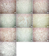
Of the 22 RCC patients with postoperative follow-up duration from 12 to 34 months (median, 19 months) who were enrolled in this study, 3 patients showed strong positive expression of Gli-1, 4 showed positive expression of ON, 3 showed positive expression of N-cadherin, 5 showed positive expression of vimentin, and 9 showed negative expression of E-cadherin, respectively. In addition, 4 patients showed negative expression of Gli-1, 1 showed negative expression of ON, 2 showed negative expression of N-cadherin, 3 showed negative expression of vimentin, and 6 showed positive expression of E-cadherin, respectively (Fig. 4).
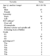
Our IHC staining of the expression of six markers and the patients' clinicopathological data showed a significant correlation between the expression of ON and renal vein sinus invasion (p=0.0218). Furthermore, the expression of vimentin had a significant correlation with lymphatic vessel invasion (p=0.0392) (Tables 1, 2).
The Hh signaling pathway functions in development. Several studies have suggested that the Hh pathway is active in RCC and promotes tumor growth, invasion, and metastasis [4]. Targeting the Hh pathway may have promising results, and Smo and Gli have been considered primary targets for anti-Hh therapeutics [20]. For example, a corn lily steroidal alkaloid, Cy, inhibits intracellular Hh signaling by blocking the activity of Smo. In this study, we demonstrated significant growth enhancements after the addition of an exogenous Shh signaling stimulator, r-Shh protein, and a growth inhibitory effect of the addition of Cy in the RenCa RCC cell line. The optimal concentration of Cy for RCC cell growth was determined to be 5 µM for this system.
EMT has recently come associate with tumor invasiveness and metastasis in RCC [21]. The mechanism may be via upregulation of mesenchymal genes such as vimentin and down-regulation of epithelial-associated markers such as E-cadherin [22]. Our in vitro mechanistic data by quantitative RT-PCR showed that even though it did not demonstrate the complete link with Shh signaling, r-Shh protein stimulated E-cadherin signaling (suppression of E-cadherin) and this was partly diminished by Cy, which may suggest a possible signaling link of Shh signaling with E-cadherin. In addition, our IHC data showed a significant correlation of vimentin with lymphatic vessel invasion (ly), which is associated with worse metastasis-free survival, disease-specific survival, and overall survival in nonmetastatic RCC patients [23].
Xu et al. [24] stated that the expression of Gli-1 is significantly up-regulated in human hepatocellular carcinoma tissue and that the expression is positively correlated with Shh signaling and vimentin. Our data for EMT up-regulation by r-Shh are supported by this study. Our human specimen data showing that vimentin expression may lead to lymphatic vessel invasion suggest that, in the link between in vitro study and the data from the human specimens, Shh signaling may relate to RCC prognosis. On the other hand, regarding the link of Hh signaling with EMT, Hh signaling mediates EMT in immature ductular cells and then contributes to fibrogenic repair in nonalcoholic fatty liver disease, and this mediation can be blocked by Cy [25]. Our study may show the potential of this link in RCC through cell study; therefore, our next task will be to study the effect of Cy in an animal study using the RCC model.
Noncanonical activation of the Hh signaling pathway is related to pancreatic cancer cell invasion and EMT through SDF-1/CXCR4 signaling [26]. Our data showed that Shh induced E-cadherin and that this induction was blocked by Cy. The IHC data also showed that a potential Shh-Gli target gene, ON [7], and vimentin were related to patients' renal vein sinus invasion and lymphatic vessel invasion, respectively, which may affect prognosis. Taken together with our results of enhanced cell proliferation by r-Shh, these data suggest that the Shh-EMT signaling link might play a role in RCC progression.
ON has several roles in different tumors, which may be attributed to the bioavailability of either the whole molecule or its cleavage products, and high ON levels are often correlated with the most aggressive and highly metastatic tumors [27]. In clinical situations, RCC often metastasizes to bone, which may cause pain, bone fracture, immobility, and decreased quality of life. Our IHC results showed that ON expression led to a significantly higher chance of renal vein sinus invasion, which is known to be related to worse prognosis [28]. Taken together, these results suggest that ON expression in RCC may show higher potential for invasion and future metastasis.
We would like to emphasize the limitations of this study. First, our sample size of patients may not have been large enough for definitive evaluation. In addition, this study lacked a mechanistic study at the protein level by use of Western blots. Third, only 2 kinds of RCC cells were tested, even though one kind of cell (ACNH cells) did not have any definite response to cyclopamine in our experimental model, and this may not be enough for definitive evaluation. These limitations will be addressed in our future projects.
Figures and Tables
 | FIG. 1
in vitro relative cell proliferation assay in renal cell carcinoma cell lines in RenCa (Panel A) and ACNH cells (Panel B) treated with dimethyl sulfoxide. Only RenCa cells showed that Cy blocked cell proliferation enhanced by r-Shh treatment. ACNH cells did not show any inhibiting effect of Cy on cell proliferation. Cell proliferation of V was set as 1.0. The treatments were performed for 48 hours. Each point represents triplicate averages±standard deviation or ON. Asterisks show significant cell proliferations compared with V. V, vehicle-treated control; S, recombinant sonic hedgehog (r-Shh) protein (1 µg/mL); Cy, cyclopamine (5 µM). |
 | FIG. 2
in vitro relative cell proliferation assay of cyclopamine (Cy) doses (0.1, 0.5, 1, 5 and 10 µM) in RenCa cells. Cy 0.5, 1, 5, and 10 µM (p=0.005, p=0015, p=0.0002, and p=0.0079, respectively) significantly inhibited cell proliferation compared with the vehicle-treated control (V), and Cy at 5 µM significantly inhibited RenCa cell growth most. Asterisks show significant inhibition of cell proliferation compared with V. |
 | FIG. 3Expression of osteonectin (ON) and epithelial-mesenchymal transition (EMT) markers (E-cadherin, N-cadherin, and vimentin) in the presence of recombinant sonic hedgehog (r-Shh) protein or cyclopamine (Cy) in RenCa cells. r-Shh protein suppressed E-cadherin (×0.266 compared with vehicle control) and this suppression was partly blocked by addition of Cy alone. However, r-Shh protein did not show induction of vimentin or ON. Asterisks show significant cell proliferation or inhibition compared with vehicle-treated control (V). S, r-Shh protein. |
 | FIG. 4Typical findings of immunohistochemical staining of renal cell carcinoma (RCC) specimens with Gli-1, osteonectin (ON), E-cadherin, N-cadherin, and vimentin antibodies are shown. Panels A and B show strong (A) and weak (B) cytoplasmic expression of Gli-1 in the RCC area (scale bars, 100.0 µm; ×200, respectively). Panels C and D show strong (C) and weak (D) cytoplasmic expression of ON in the RCC area. (scale bars, 100.0 µm; ×200, respectively). Panels E and F showed strong (E) and weak (F) membrane expression of E-cadherin in the RCC area (scale bars, 100.0 µm; ×200, respectively). Panels G and H show strong (G) and weak (H) membrane expression of N-cadherin in the RCC area (scale bars, 100.0 µm; ×200, respectively). Panels I and J show strong (I) and weak (J) cytoplasmic expression of vimentin in the RCC area (scale bars, 100.0 µm; ×200, respectively). |
References
1. Cho IC, Chung J. Current status of targeted therapy for advanced renal cell carcinoma. Korean J Urol. 2012. 53:217–228.
2. Sanchez P, Hernandez AM, Stecca B, Kahler AJ, DeGueme AM, Barrett A, et al. Inhibition of prostate cancer proliferation by interference with SONIC HEDGEHOG-GLI1 signaling. Proc Natl Acad Sci U S A. 2004. 101:12561–12566.
3. Datta S, Datta MW. Sonic Hedgehog signaling in advanced prostate cancer. Cell Mol Life Sci. 2006. 63:435–448.
4. Dormoy V, Danilin S, Lindner V, Thomas L, Rothhut S, Coquard C, et al. The sonic hedgehog signaling pathway is reactivated in human renal cell carcinoma and plays orchestral role in tumor growth. Mol Cancer. 2009. 8:123.
5. Li N, Felber K, Elks P, Croucher P, Roehl HH. Tracking gene expression during zebrafish osteoblast differentiation. Dev Dyn. 2009. 238:459–466.
6. Guweidhi A, Kleeff J, Adwan H, Giese NA, Wente MN, Giese T, et al. Osteonectin influences growth and invasion of pancreatic cancer cells. Ann Surg. 2005. 242:224–234.
7. Shigemura K, Huang WC, Li X, Zhau HE, Zhu G, Gotoh A, et al. Active sonic hedgehog signaling between androgen independent human prostate cancer cells and normal/benign but not cancer-associated prostate stromal cells. Prostate. 2011. 71:1711–1722.
8. Sethi S, Macoska J, Chen W, Sarkar FH. Molecular signature of epithelial-mesenchymal transition (EMT) in human prostate cancer bone metastasis. Am J Transl Res. 2010. 3:90–99.
9. Rhim AD, Mirek ET, Aiello NM, Maitra A, Bailey JM, McAllister F, et al. EMT and dissemination precede pancreatic tumor formation. Cell. 2012. 148:349–361.
10. Lim M, Chuong CM, Roy-Burman P. PI3K, Erk signaling in BMP7-induced epithelial-mesenchymal transition (EMT) of PC-3 prostate cancer cells in 2- and 3-dimensional cultures. Horm Cancer. 2011. 2:298–309.
11. Xu X, Zhou Y, Xie C, Wei SM, Gan H, He S, et al. Genome-wide screening reveals an EMT molecular network mediated by Sonic hedgehog-Gli1 signaling in pancreatic cancer cells. PLoS One. 2012. 7:e43119.
12. Joost S, Almada LL, Rohnalter V, Holz PS, Vrabel AM, Fernandez-Barrena MG, et al. GLI1 inhibition promotes epithelial-to-mesenchymal transition in pancreatic cancer cells. Cancer Res. 2012. 72:88–99.
13. Onishi T, Ogawa T, Hayashibara T, Hoshino T, Okawa R, Ooshima T. Hyper-expression of osteocalcin mRNA in odontoblasts of Hyp mice. J Dent Res. 2005. 84:84–88.
14. Matsuoka TA, Kaneto H, Stein R, Miyatsuka T, Kawamori D, Henderson E, et al. MafA regulates expression of genes important to islet beta-cell function. Mol Endocrinol. 2007. 21:2764–2774.
15. Aoi J, Endo M, Kadomatsu T, Miyata K, Nakano M, Horiguchi H, et al. Angiopoietin-like protein 2 is an important facilitator of inflammatory carcinogenesis and metastasis. Cancer Res. 2011. 71:7502–7512.
16. Kurahashi T, Miyake H, Hara I, Fujisawa M. Expression of major heat shock proteins in prostate cancer: correlation with clinicopathological outcomes in patients undergoing radical prostatectomy. J Urol. 2007. 177:757–761.
17. Oue T, Yoneda A, Uehara S, Yamanaka H, Fukuzawa M. Increased expression of the hedgehog signaling pathway in pediatric solid malignancies. J Pediatr Surg. 2010. 45:387–392.
18. Bozkurt SU, Ayan E, Bolukbasi F, Elmaci I, Pamir N, Sav A. Immunohistochemical expression of SPARC is correlated with recurrence, survival and malignant potential in meningiomas. APMIS. 2009. 117:651–659.
19. Koo JS, Jung W. Clinicopathlogic and immunohistochemical characteristics of triple negative invasive lobular carcinoma. Yonsei Med J. 2011. 52:89–97.
20. Lauth M, Toftgard R. The Hedgehog pathway as a drug target in cancer therapy. Curr Opin Investig Drugs. 2007. 8:457–461.
21. Bostrom AK, Moller C, Nilsson E, Elfving P, Axelson H, Johansson ME. Sarcomatoid conversion of clear cell renal cell carcinoma in relation to epithelial-to-mesenchymal transition. Hum Pathol. 2012. 43:708–719.
22. Barnett P, Arnold RS, Mezencev R, Chung LW, Zayzafoon M, Odero-Marah V. Snail-mediated regulation of reactive oxygen species in ARCaP human prostate cancer cells. Biochem Biophys Res Commun. 2011. 404:34–39.
23. Katz MD, Serrano MF, Humphrey PA, Grubb RL 3rd, Skolarus TA, Gao F, et al. The role of lymphovascular space invasion in renal cell carcinoma as a prognostic marker of survival after curative resection. Urol Oncol. 2011. 29:738–744.
24. Xu QR, Zheng X, Zan XF, Yao YM, Yang W, Liu QG. Gli1 expression and its relationship with the expression of Shh, Vimentin and E-cadherin in human hepatocellular carcinoma. Xi Bao Yu Fen Zi Mian Yi Xue Za Zhi. 2012. 28:536–539.
25. Syn WK, Jung Y, Omenetti A, Abdelmalek M, Guy CD, Yang L, et al. Hedgehog-mediated epithelial-to-mesenchymal transition and fibrogenic repair in nonalcoholic fatty liver disease. Gastroenterology. 2009. 137:1478–1488.e8.
26. Li X, Ma Q, Xu Q, Liu H, Lei J, Duan W, et al. SDF-1/CXCR4 signaling induces pancreatic cancer cell invasion and epithelial-mesenchymal transition in vitro through non-canonical activation of Hedgehog pathway. Cancer Lett. 2012. 322:169–176.
27. Podhajcer OL, Benedetti L, Girotti MR, Prada F, Salvatierra E, Llera AS. The role of the matricellular protein SPARC in the dynamic interaction between the tumor and the host. Cancer Metastasis Rev. 2008. 27:523–537.
28. Bedke J, Buse S, Pritsch M, Macher-Goeppinger S, Schirmacher P, Haferkamp A, et al. Perinephric and renal sinus fat infiltration in pT3a renal cell carcinoma: possible prognostic differences. BJU Int. 2009. 103:1349–1354.


 PDF
PDF ePub
ePub Citation
 Print
Print


 XML Download