

 PDF
PDF ePub
ePub Citation
 Print
Print


INTRODUCTION
After the first reported shock wave lithotripsy (SWL) of renal calculi in 1980 by Schmiedt and Chaussy [1], SWL has been improved and has gained acceptance as a first-line treatment option for small urinary stones. European Association of Urology guidelines recommend SWL as a first choice for a stone <1 cm in size located in the kidney. Compared with flexible ureteroscopy and percutaneous nephrolithotomy, SWL can be performed in outpatient clinics, and the patient does not require general anesthesia. However, despite the advantages of SWL, pain that results from shock wave treatment is still commonly bothersome to patients and may be one of the reasons some patients hesitate to choose SWL. Furthermore, pain perception during SWL could cause patients to change body position, which could disturb the therapeutic shock wave focus and adversely affect the SWL success rate.
Following improvements in SWL technology, analgesic requirements for pain control during SWL have dramatically decreased [2]. Furthermore, several studies have shown that analgesics such as opioids, nonsteroidal anti-inflammatory drugs, and anesthetic topical creams provide adequate pain control [3-5]. Several clinical factors such as sex, age, body mass index (BMI), and stone location have been elucidated as predictive factors for SWL-related pain [6-8]. However, little clinical data is available regarding patient positioning during SWL. The aim of this study was to evaluate the association between SWL-related pain and patient position (supine and lateral) during SWL.
MATERIALS AND METHODS
1. Patients and methods
We retrospectively reviewed the medical records of patients who were treated with SWL for renal stones between May 2010 and August 2011 at Samsung Medical Center. Patients eligible for inclusion in this study were those who were diagnosed with a single renal stone smaller than 20 mm by noncontrast computed tomography (CT) before treatment and had undergone their first session of SWL.
A total of 162 patients were eligible for this study and were evaluated. Patient demographics, pain scores during SWL, and stone characteristics including size, location, stone-free rate, success rate, and skin-to-stone distance (SSD) were reviewed. We measured the size of the stone at its maximal diameter and categorized the location of the stones into one of three areas: upper, mid, or lower portion. The longitudinal borders were defined as axial polar lines drawn at the level of the medial lip of the renal cortical parenchyma. When calculating SSD, we used the methodology of Pareek et al. [9] that is based on noncontrast CT. All patients were evaluated 4 weeks after the first SWL session by use of KUB or renal ultrasound to assess the presence of remnant renal stones. Successful SWL was defined as complete stone clearance or presence of residual stone fragments <3 mm, whereas failure was defined as the presence of residual fragments larger than 3 mm.
2. Shock wave lithotripsy procedure
All SWL treatments were performed by using a MODULITH SLX-F2 system (STORZ Medical AG, Tägerwilen, Switzerland), which has an electromagnetic cylinder unit that permits the use of X-ray or ultrasound for stone focusing. This lithotripter has a cylindrical shock wave source under the radiotransparent patient table. Thus, fluoroscopic projections and localization were done by using anteroposterior and lateral directions according to the known standards of usual form. In addition, this system has a coaxial (in-line) arrangement of the diagnostic ultrasound transducer and therapeutic shock wave source, which ensures that shock waves and ultrasound waves travel through the same regions so that only minimal deviations occur regarding their propagation.
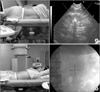
If stone targeting in the supine position on fluoroscopy was possible, we treated the patient in the supine position. However, if the stone was not visualized well in the supine position upon fluoroscopy, the patient was treated in the lateral position under ultrasound guidance (Fig. 1).
Patients were asked if pain control would be necessary during the procedure, but medication was not administered before SWL. When patients asked for pain control, a nonsteroidal anti-inflammatory drug (30 mg/mL; Ketorolac Tromethamine, Hana Pharm Co., Seoul, Korea) was injected intramuscularly during SWL.
All patients were treated with up to 3,000 shock waves, with a maximum power of 12.5 kV at 90 shocks per minute. Immediately after the procedure, the degree of pain was evaluated by using a 10-point visual analogue scale (VAS), and both the average degree of pain (VAS-avg) and maximum pain (VAS-max) were assessed together. If the patient received an intramuscular injection for pain control, we assessed both the maximum and average degrees of pain perception in the same manner before injection.
3. Statistical analysis
A total of 162 patients were divided into two groups according to position during SWL. One hundred thirteen patients were treated in the supine position and 49 patients were treated in the lateral position. To obtain an unbiased estimation of the positional effect on pain severity during SWL, we matched patients between groups at a ratio of 1:1 without knowing the pain scores or success results. A patient in the lateral group was matched with a patient in the supine group according to sex, age (±2 years), BMI (±2 units), location of stone (either same or adjacent location in kidney, e.g., lower and mid portion or upper and mid portion could be matched), and stone size (±2 mm). Only matched patients were selected for review, and each group had 34 patients.
All statistical analyses were performed by using the IBM SPSS ver. 18.0 (IBM Co., Armonk, NY, USA). Group differences were evaluated by using Pearson chi-square test and Student t-test for categorical data and continuous data, respectively. Statistical significance was defined as p<0.05.
RESULTS
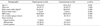
The mean age of the patients was 53.3±12.9 years. The associated demographic data are presented in Table 1. After matching, groups did not demonstrate significant differences in terms of sex, age, BMI, SSD, renal stone size, or renal stone location, except for the radio-opacity of stones. In the supine group, all patients had a radio-opaque stone. In the lateral group, however, only 16 of the 34 patients (47.1%) had a radio-opaque stone and the difference between groups was statistically significant (p<0.01).
The comparison of pain scores during the SWL procedure between the two groups is presented in Table 2. Pain scores during the procedures were affected by patient position, for which statistically significant differences were demonstrated. The mean VAS-avg of the supine and lateral groups was 3.1±1.7 and 1.2±1.0, respectively (p<0.05). The VAS-max of the lateral position group was also significantly lower than that of the supine position group (2.5±1.8 vs. 4.7±1.9, p<0.05). Most patients tolerated the procedure well without the use of analgesics. Of the patients in the lateral position, only two (5.9%) requested medication for pain control, compared with six patients (17.6%) in the supine group. However, there were no significant differences in the usage of analgesics, and no side effects of analgesics were observed.
In the subgroup analysis, we compared VAS-avg and VAS-max in patients who had radio-opaque stones. The supine group still suffered more severe pain (Table 3). The VAS-avg of the lateral and supine groups was 1.2±1.0 and 3.1±1.7, respectively (p<0.001). VAS-max of the lateral group (2.7±2.0) was lower than that of the supine group (4.7±1.9, p=0.02).
The success rate and stone-free rate of both groups are summarized in Table 4. The proportions of patients who were stone-free after the first session of SWL within the supine and lateral groups were 32.4% and 26.5%, respectively (p=0.597). In addition, the overall success rate of the supine group (47.1%) was higher than that of the lateral group (38.2%), but this difference was not statistically significant (p=0.465).
No major complications such as perirenal hematoma or urinary tract infection occurred, and none of the patients required an emergency room visit after SWL.
DISCUSSION
Currently, extracorporeal SWL is a first-line treatment option for small renal stones. Pain control during SWL has been an important issue to date, because pain during the procedure may result in patient withdrawal and may decrease the success rate of the procedure.
Several studies have investigated the relationships of various factors with pain perception during SWL treatment. Tokgoz et al. [7] suggested that pain during SWL may be better tolerated in males than in females and that the first session of SWL is typically more comfortable for patients than subsequent sessions. Oh et al. [6] also reported that the subjective pain score was impacted by patient age, sex, and the location of the stone, but it was not affected by the size or laterality of the stone.
In practice, we observed that patients in the lateral position during SWL complained of pain less frequently than did patients in the supine position. However, the effects of body position on pain severity during SWL have not been well studied previously. Thus, we decided to investigate the relationship between body position and pain severity during SWL.
To minimize the potential influence of confounding factors such as sex, age, BMI, stone location, and stone size, we designed a 1:1, pair-matched, case-control study. Moreover, we separately recorded the maximum and average VAS pain scores during the procedure. The VAS is a simple and widely used method to assess variations in pain intensity [10]. The perception of pain is a subjective response, and the intensity and quality of the pain may be affected by various environmental factors [11,12]. Thus, if we only assessed the maximum pain severity during SWL, the reliability of our results may have been compromised. By recording both the average and maximum pain severity, we attempted to obtain a more objective evaluation of pain during SWL
In the results of our study, the patients who were treated in the lateral position had a significantly lower perception of pain than did those who were treated in the supine position by use of both the maximum and average VAS score (Table 2). It is difficult to precisely interpret the reason behind our results, but we surmise that different shock wave entry points owing to different patient positions are an important factor.
Vergnolles et al. [13] analyzed predictive risk factors of pain severity during SWL. Their findings demonstrated that the presence of a "rib projected stone" was a significant independent risk factor for pain. They defined "rib projection" as when the stone was close to the rib as observed via fluoroscopy and assumed the reason for the pain was probably because of periosteum sensitivity and irritation of the intercostal nerve. This study demonstrated that in addition to patient demographics, the course that the shockwave travels is also related to the amount of pain. In the current study, the different shock wave entry points resulted in different shockwave pathways; consequently, this created various patterns of nociceptive stimulation.
The proportion of radio-opaque stones differed between the two groups, which is a shortcoming of this study. However, the number of radio-opaque stones differed significantly between the two groups because radiolucent stones could not be visualized in the supine position by ultrasound by use of the MODULITH SLX-F2 system, which has an ultrasound probe beneath the table. However, at the beginning of the current study, it was assumed that the radio-opacity of a stone would not affect pain severity during SWL. Therefore, we did not consider the stone radio-opacity as a matching variable. We also compared pain severity only in patients with radio-opaque stones in both groups and the results did not demonstrate a statistically significant correlation between pain scores and stone opacity (Table 3).
To our knowledge, these results are the first clinical data to indicate that body position may play a significant role in pain severity during SWL. Thus, we think that the current study is valuable in providing information about pain reduction during SWL.
Potential limitations of the current study include the small sample size and retrospective design. A clearer idea of the differences between the two positional groups during SWL could be formed if a prospective study with a larger sample size was conducted. Currently, we are planning a prospective trial to address this issue. Although the design of this study was retrospective, all data were recorded in a prospective manner, in which the investigators did not know the study design, and we achieved two similar patient groups by matching patients according to potential confounding factors for pain perception during SWL. Another limitation is that we cannot ensure that the same results would be obtained by use of lithotripters other than the MODULITH SLX-F2 lithotripter.
CONCLUSIONS
The results of our study showed that the mean VAS-avg and VAS-max of the lateral group were significantly lower than those of the supine group. Moreover, analgesic usage tended to be lower in the lateral group, although there was no statistical significance to this observation. The present findings indicate that patients with solitary renal stones suffer more SWL-related pain in the supine position than in the lateral position. Therefore, we suggest that positioning of patients during SWL should be considered a predictive factor for SWL-related pain.


 XML Download