

 PDF
PDF ePub
ePub Citation
 Print
Print


INTRODUCTION
Magnetic resonance imaging (MRI) for prostate cancer (PC) diagnosis was previously mainly performed with conventional T2-weighted imaging (WI), and the quality for this purpose is in general limited to staging for organ-confined PC or the presence of extracapsular extension and seminal vesicle invasion. Recently, diffusion-weighted imaging (DWI) MRI has become more common and may expand the diagnostic role of MRI in PC by providing more specific information regarding tumor location, size, and aggressiveness [1-7].
The Gleason score (GS) is a well-known indicator of PC aggressiveness. One of the most important goals in radical prostatectomy for PC is the avoidance of PC recurrence, and the GS, surgical margin status, and capsule invasion are commonly used to assess the probability of PC recurrence [8-10]. Prostate biopsy is another important diagnostic modality for determining the GS in PC diagnosis. In addition, Woodfield et al. [1] found that DWI MRI had some efficacy for the discrimination of low, intermediate, and high-risk PC by use of prostate biopsy specimens, and Hambrock et al. [11] reported that apparent diffusion coefficients (ADCs) at 3.0 T showed an inverse relationship to GS in peripheral zone PC by using prostatectomy specimens. However, debate continues about differences in the results from prostate biopsy and prostatectomy specimens [12]. We as urologists know the limitations of prostate biopsy specimens for predicting PC recurrence.
In this study, we compared DWI MRI data and GS findings from prostatectomy specimens. Because a high GS apparently leads to a higher PC recurrence ratio with high malignant potential, such preoperative findings may be informative for detailed decision making concerning prostatectomy, especially in cases with the prediction of a high GS from DWI findings.
MATERIALS AND METHODS
1. Patients
Between January 2009 and October 2011, 105 consecutive men with full data for analyses and who did not receive neoadjuvant hormonal therapy were evaluated in this study. Full data included age, preoperative serum prostate-specific antigen (PSA) values, prostate MRI data including DWI, and pathological GS, margin status, and capsule invasion as shown by prostatectomy specimens. All cases underwent radical retropubic prostatectomy performed via the antegrade approach with the inclusion of intrapelvic lymphadenectomy.
2. MRI testing and technique
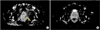
All prostate MRI examinations were performed before prostate biopsy for patients suspected of having PC by use of an Achieva 1.5-T A-series MRI system (Philips Medical Systems, Einthoven, Netherlands). The MRI protocol included an axial T1-WI and axial, sagittal, and coronal T2-WI, DWI, and ADC map. The B-factor set in this study for DWI MRI was 2000. The criterion for positive cancer in DWI MRI in this study was defined as low-intensity imaging on the ADC map and this area was defined as a PC-positive lesion in this study [13]. MRI diagnosis was performed mainly by an experienced radiologist (18 years of practice after board certification).
3. Analyses of DWI MRI and PC
We analyzed the relationship between PC-positive findings in DWI MRI and the patients' pathological data such as GS, margin status, capsule invasion, and pathological T stage (pT). In addition, in 62 patients with PC-positive DWI MRI findings, we compared the relationship between tumor size measured by DWI MRI and GS in 55 patients with a single DWI MRI PC-positive lesion. Moreover, we compared the GS of patients with multiple tumor lesions found by DWI MRI with that of patients with a single DWI MRI PC-positive lesion. We defined GS≥4+3 as a high GS.
4. Statistical analysis
Statistical analyses were performed by using Student's t-test or chi-square test for univariate analysis with p<0.05 considered to indicate statistical significance. Multivariate analyses were performed by multiple regression analysis with a p-value of <0.05 and partial regression coefficient of >0.5 to indicate statistical significance of the regression coefficient. These tests were performed with JSTAT (Java Virtual Machine Statistics Monitoring Tool, Oracle Co., Redwood City, CA, USA).
RESULTS
1. Patients and pathological data
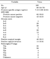
In this study, 105 consecutive men (median age, 68 years; range, 54 to 76 years) with full DWI MRI and clinical and pathological data who underwent radical prostatectomy for PC were evaluated. Detailed patient backgrounds are shown in Table 1. The median PSA level was 7.10 ng/mL (range, 1.90 to 40.61 ng/mL). In addition, 40 patients (38.1%) had positive surgical margin and 11 (10.5%) had positive capsule invasion. The prostatectomy specimens showed 1 patient (0.95%) with GS 3+2, 29 patients (27.6%) with GS 3+3, 38 patients (36.2%) with GS 3+4, 1 patient (0.95%) with GS 3+5, 22 patients (21.0%) with GS 4+3, 12 patients (11.4%) with GS 4+4, 1 patient (0.95%) with GS 4+5, and 1 patient (0.95%) with GS 5+4 (Table 1).
2. DWI MRI
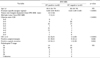
The DWI MRI findings showed that 62 of 105 patients had PC-positive DWI MRI results, whereas 43 had PC-negative DWI MRI results (Table 1 and Fig. 1). In addition, in the 62 patients with PC-positive DWI MRI results, the longest diameter for tumor size was 12 mm (range, 4 to 24 mm). The relationship between tumor size measured by DWI MRI and GS in 55 patients with a single DWI MRI PC-positive lesion was not significant (p>0.05). However, in the comparison of GS between patients with multiple tumor lesions (the number of patients with multiple tumor lesions by DWI MRI was 7) by DWI MRI and those with a single tumor lesion (n=55), we found that the patients with a single tumor lesion had significantly higher GSs than did those with multiple tumor lesions (p=0.0301) (Table 2). In addition, regarding localized or locally advanced PC, we observed that cases with locally advanced PC had a significantly higher ratio of positive DWI MRI findings (p=0.0059). Regarding GS distribution according to localized or locally advanced PC, we demonstrated that the cases with locally advanced PC had a significantly higher ratio of a high GS (8 or more, p=0.0051) (Table 3).
3. Statistical data
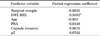
The univariate analysis showed that the PC-positive DWI MRI group (n=62) had a significantly higher PSA (p<0.0001), higher GS (p<0.0001), higher ratio of positive capsule invasion (p=0.0026), higher ratio of positive surgical margins (p=0.0027), and higher pT stage (p=0.0020) than did the PC-negative DWI MRI group (n=43) (Table 2). The multiple regression analysis with DWI MRI as the response variable and other factors (GS, age, PSA, positive surgical margins, capsule invasion, and pT) as predictor variables showed that the PC-positive DWI MRI group (n=62) had significantly higher GSs and a higher ratio of positive surgical margins than did the PC-negative DWI MRI group (n=43). However, on the contrary, the multiple regression analysis with positive surgical margins as the response variable and other factors (DWI MRI, age, PSA, GS, capsule invasion, and pT) as predictor variables showed no significant application (p=0.0665). On the other hand, importantly, multiple regression analyses with GS as the response variable and other factors (DWI MRI, age, PSA, positive surgical margins, capsule invasion, and pT) as predictor variables showed a statistically significant application (p<0.0001), and DWI MRI was a significant predictor variable for GS (partial regression coefficient, 0.5085). Taken together, these results suggest that DWI MRI and GS, not positive surgical margin, showed a significant relationship in both directions (Table 4).
DISCUSSION
PC is the second most common male cancer in the United States. Diagnostic options are varied and include many choices [8]. With regard to imaging for detection of PC, the increasing exploitation of MRI has been remarkable; in particular, T2-WI has been widely used for PC diagnosis [14]. In addition, T2-WI MRI has been used to identify prostate zonal anatomy and extracapsular integrity for PC diagnosis [15]. The combination of conventional T2-WI MRI with other MRI modalities is noted to offer improved diagnostic performance for PC. This preoperative diagnosis is also important for decision making regarding surgical margins and neurovascular bundle preservation [8,9,15] as mentioned above.
PC recurrence is often seen after local therapy such as surgery or radiation [16], and its risk factors are well-known [17]. Therefore, treatments are recommended according to guidelines or risk criteria such as D'Amico's classification for standardizing therapeutic strategies for PC. Serum PSA, T stage, and GSs are well recognized to predict the risks for PC recurrence. In particular, a GS of 4+4 or higher is considered an element of high-risk PC, and there is definitely an apparent difference in malignancy between GS 4+3 and 3+4 [18]. On the other hand, specimens from prostate biopsies are small, and the PC will occupy much less volume in one prostate biopsy sample in most cases of organ-confined PC. This may be the cause of the discrepancy noted above between GSs based on biopsy samples versus prostatectomy specimens [19]. This study investigated how the GS could be assumed preoperatively by using DWI MRI, which is a comparatively new tool, with T1- or T2-WI to assess the risk for PC recurrence and the extent of malignancies and also for intraoperative decision making regarding surgical margins and neurovascular bundle preservation [14]. Several studies have reported that PC location in the peripheral zone could be detected by DWI MRI over T2-WI MRI and that combined DWI and T2-WI MRI offers better sensitivities and specificities than does DWI MRI [20]. Bittencourt et al. [8] showed that the GS from prostatectomy specimens rather than prostate biopsy specimens correlated well with DWI MRI findings. Our data showed a significant correlation between GSs of 4+3 or higher and the DWI MRI results, which agrees with their data.
Moreover, our data also showed that serum PSA was significantly higher in the PC-positive DWI MRI group than in the PC-negative DWI MRI group; however, serum PSA levels, especially low PSA levels, do not necessarily reflect or predict prognoses and PC recurrence differently from, for instance, PSA velocity [21]. Therefore, our investigated categories, such as the GS, may reflect the extent of cancer aggressiveness or the risk of PC recurrence. As such, the relations between these variables and PC-positive findings in DWI MRI may be considered with significance. In this situation, we should consider the correlation of tumor size or single or multiple tumors from the DWI MRI findings with the GS. We found that patients with single tumors had significantly higher GSs than did patients with multiple tumors. This finding might contribute to the prediction of higher GSs. In particular, as to single or multiple tumors and their correlation with GSs, we assume that the single tumors detected by DWI MRI tended to be larger than the multiple tumors and thus the former may have had higher malignant potential with the characteristics of tumor spreading or invasion.
Prostatectomy is performed for localized or locally advanced PC in general. Our data showed that locally advanced PC was associated with a significantly higher ratio of PC-positive DWI MRI findings (p=0.0059) and a high GS (8 or more) (p=0.0051), which suggests that PC-positive DWI MRI may reveal PC with a high malignant potential.
There are some limitations to this research study. First, this was a retrospective study in which several cases were excluded owing to insufficient data for analysis, and the total number of cases might not be high enough to draw definitive conclusions. Next, the low-intensity findings were subjectively diagnosed by radiologists. As mentioned above, several previous reports offered calculated ADC values and objective evaluations [8,22,23]. Our next task should be an expanded study using quantified data with a greater number of patients, and future research should focus on linking low-intensity DWI MRI with pathological findings in terms of cancer location and GS. In addition, we could not evaluate the significance of PC-positive DWI MRI results according to equivalent tumor volume because of insufficient pathological data. This should be our next goal to accomplish.
CONCLUSIONS
Our findings using low-intensity DWI MRI significantly correlated with analysis based on prostatectomy specimens to diagnose PC with more malignant potential (GS≥4+3). Our next aim is a longer duration follow-up study to determine whether PC recurrence is ultimately higher in cases with positive findings based on low-intensity DWI MRI.

 XML Download