

 PDF
PDF ePub
ePub Citation
 Print
Print


INTRODUCTION
According to GLOBOCAN 2008 reported by Ferlay et al. [1], bladder cancer incidence ranked fourth and bladder cancer mortality ranked seventh in developed countries. Although bladder cancer can be treated with surgery, intravesical chemotherapy, and systemic chemotherapy in most cases, it is hard to delay the progression of the cancer despite an appropriate treatment strategy. In particular, superficial bladder cancer accounts for 70% of the total cases of bladder cancer and tends to progress to muscle-invasive cancer even after proper treatment [2,3].
Zinc has antioxidant, antiinflammatory, and proapoptotic activity and plays a role in genetic stability and function [4,5]. Studies in an ovarian cancer cell line and choriocarcinoma cells have suggested that zinc can induce the apoptosis of cancer cells [6,7]. A study to determine the apoptogenic effect of zinc through a high difference in concentration between normal and prostate cancer cell lines was also conducted [8]. Citrate, one of the components of the Krebs cycle, has been reported to elevate the solubility of heavy metals by binding to heavy metal ions, and upon binding with zinc it may increase the bioavailability of zinc [9].
In the present study, we aimed to demonstrate the antiproliferative activity of a zinc-citrate compound in a bladder cancer cell line and to investigate its inhibitory mechanism of action.
MATERIALS AND METHODS
1. Preparation of zinc-citrate compound
The zinc-citrate compound was composed of zinc chloride (Sigma-Aldrich Co., St. Louis, MO, USA) and citric acid anhydrous (Sigma-Aldrich Co.). The compound was boiled for 15 minutes in a 121℃ autoclave.
2. Cell culture
The bladder cancer cell line MBT-2 (American Type Culture Collection, Manassas, VA, USA) was cultured in Rosewell Park Memorial Institute (RPMI) 1640 supplemented with 10% fetal bovine serum (FBS), L-glutamine (300 mg/l), 25 mM 4-(2-hydroxyethyl)-1-piperazineethanesulfonic acid, and 25 mM NaHCO3.
3. Intracellular zinc concentration
The intracellular zinc concentration was measured with the Zinc Assay Kit (Sentinel CH, Milan, Italy). MBT-2 cells were treated with normal saline or zinc-citrate compound for 1, 3, and 6 hours. Cells were centrifuged at 1,500 rpm for 5 minutes. Cells (1×10⁶/100 µl in phosphate buffered saline [PBS]) were sonicated three times for 10 seconds and centrifuged at 14,000 rpm. Solution AC was prepared with 40 mg of Reagent C and 1 vial of Reagent A. A total of 5 µl of sample was mixed with 100 µl of solution AC and 10 µl of Reagent C and was read in a Bio-Rad Model 3550 Microplate Reader (Richmond, CA, USA).
4. Cell viability assay
MBT-2 cells were treated at different time intervals and with various doses to evaluate the effects of the zinc-citrate compound on cell growth and survival. Cells were inoculated into 96-well plates at a density of 1×10⁴ cells/well for 4 hours. Subsequently, cells were cultured in medium containing diverse concentrations of normal saline and zinc-citrate compound (0.25/5, 0.5/10, 1/20, and 2/40 mM) for 24 hours. The IC50 (the half maximal inhibitory concentration) was obtained, and cells were incubated with zinc-citrate compound at the IC50 concentration for 6, 12, 24, and 48 hours. After exposure to the zinc-citrate compound, 20 µl CCK solution (1 mg/ml in PBS) was added to each well, and the plates were incubated at 37℃ for 4 hours The plates were read at a wavelength of 450 nm by using the Bio-Rad Model 3550 Microplate Reader (Richmond, CA, USA). The wells containing only RPMI 1640-FBS and CCK were used as the control group.
5. Mitochondrial aconitase activity
Mitochondrial (m)-aconitase activity in the cell extracts was measured by using the Bioxytech aconitase-340 assay (Oxis Research, Foster City, CA, USA). MBT-2 cells were treated with the zinc-citrate compound for 1, 2, and 4 hours and were observed at a wavelength of 340 nm for 5 minutes at 37℃.
6. DNA laddering analysis
MBT-2 cells (2×10⁶) treated with the zinc-citrate compound (0.4/8 mM) for 6, 12, 24, or 48 hours were washed with PBS. An amount of 200 µl binding/lysis buffer was added and the total volume was adjusted to 400 µl and the samples were mixed. After the samples were incubated at 15 to 25℃ for 10 minutes, 100 µl isopropanol was added and the samples were shaken. To a filter tube, a collection tube was attached and subsequently the samples were added to the filter tube and centrifuged at 800 rpm for 1 minute. Then, 500 µl washing buffer was added and the tubes were centrifuged at 800 rpm for 1 minute twice. The samples were then centrifuged a third time at 13,000 rpm for 10 seconds. An amount of 100 µl elution buffer was added, and the samples were centrifuged at 800 rpm for 1 minute. Finally, electrophoresis was performed on 1% agarose gel at 75 V for 1.5 hours, and the results were assessed under ultraviolet light.
7. Western blot analysis
MBT-2 cells treated with the zinc-citrate compounds for 6, 12, 24, or 48 hours were extracted by centrifuging at 4℃, 2,000 rpm, for 5 minutes. The reaction was lysed by the addition of 100 µl lysis buffer for 30 minutes at room temperature. MBT-2 cell lysate exposed to the zinc-citrate compound (50 µg of protein) was electrophoresed on 10% sodium dodecyl sulfate/polyacrylamide gels at 100 V for 2 hours and were treated for 1 hour with PBS containing 5% nonfat milk and 0.05% Tween 20. p53 and p21waf1 were assessed, and the antiapoptotic protein Bcl-2 as well as Bcl-xL and the proapoptotic protein Bax were assessed. The samples were reacted with the primary antibody to p53, p21waf1, Bcl-2, Bcl-xL, and Bax (diluted 1:500) at 4℃ overnight. Membranes were washed 3 times with PBS containing 0.05% Tween 20 and reacted with anti-mouse or anti-rabbit secondary antibody (Calbiochem, San Diego, CA, USA) diluted 1:5,000 for 1 hour at room temperature. Each membrane was washed with PBS containing 0.05% Tween 20 three times. Specific protein bands were examined by using the enhanced chemiluminescence Western blotting system (Amersham, Piscataway, NJ, USA).
8. Caspase-3 activity assay
MBT-2 cells exposed to the zinc-citrate compound were washed with PBS, 50 µl lysis buffer was added, and the cells were kept on ice for 10 minutes. The cell lysate was centrifuged at 4℃ and 12,000 rpm and the supernatant was transferred to a new tube and stored on ice. Caspase-3 activity was measured with a Caspase-3 colorimetric protease assay kit (BioSource, Camarillo, CA, USA). Then 50 µl lysis buffer was adjusted to contain 100 µg of protein, and 10 µl of 0.1 M dithiothreitol (DTT) was mixed with 1 ml of 2× reaction buffer. Next, 50 µl of 0.1 M DTT-2× reaction buffer and 5 µl of 4 mM DEVD-pNA was added to the samples. The samples were then wrapped with aluminum foil and kept at 37℃ for 2 hours. Absorbance was measured at 405 nm by using an enzyme-linked immunosorbent assay plate reader.
9. Statistical analysis
All experiments were performed on three separate cultures. All data are presented as the mean±standard deviation, where p<0.05 was considered to be statistically significant. Overall comparisons between groups were performed by using SAS 8.0 (SAS Institute Inc., Cary, NC, USA). Repeated-measures analysis of variance with Duncan's test was performed to detect differences over the time course and between the groups.
RESULTS
1. Intracellular zinc concentration
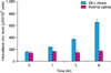
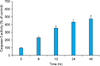
The zinc concentration in the MBT-2 cells before the addition of the zinc-citrate compound was low. The intracellular zinc concentration was recorded as 162±12 µM/l/106 cells, 238±4 µM/l/106 cells, 370±12 µM/l/106 cells, and 657±47 µM/l/106 cells at 0, 1, 3, and 6 hours after the treatment and the concentration became higher with the passage of time. However, the zinc concentration in the group treated with normal saline was not changed (Fig. 1).
2. Cell viability assay
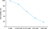
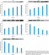
MBT2 cells exposed to the zinc-citrate compound showed a pattern of decrease in a time- and dose-dependent manner. MBT2 cells exposed to the diverse concentrations of the zinc-citrate compound for 24 hours showed a clear reduction of cell viability in the 3-(4,5-Dimethylthiazol-2-yl)-2,5-diphenyltetrazolium bromide assay (p<0.0001).
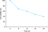
The number of cells was reduced by around 24.7% and 93.5% after treatment with the 0.25/5 mM and 2/40 mM zinc/citrate compounds, respectively. Thus, the antiproliferative effect in MBT-2 was increased at a higher concentration. The IC50 of the zinc-citrate compound against MBT2 was estimated to be 0.5/10 mM. At the IC50, the antiproliferative effect of the zinc citrate compound was raised with longer exposure time (Figs. 2, 3).
3. Mitochondrial aconitase activity
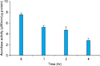
M-aconitase activity was reduced significantly from 7.4±0.3 µM/min/µg protein to 2.8±0.4 µM/min/µg protein at 4 hours after the treatment (Fig. 4).
4. DNA laddering analysis
To characterize the mechanism of cell death, DNA laddering analysis was performed. The migration of fragmented DNA as a laddering pattern was observed by electrophoresis in MBT2 cells. Apoptosis of MBT-2 was found from the results of DNA laddering.
5. Western blot
The expression of p53 and p21waf1 was elevated in the MBT2 cells when the zinc-citrate compound was added. The expression of the antiapoptotic proteins Bcl-2 and Bcl-xL was reduced, and simultaneously, the expression of the proapoptotic protein Bax was elevated and the Bcl-2/Bax ratio and the Bcl-xL/Bax ratio were decreased with time (Fig. 5).
The zinc-citrate compound enhanced the expression of p21waf1 and p53, suppressed the expression of Bcl-2 and Bcl-xL proteins, and stimulated the expression of Bax protein.
6. Caspase-3 activity
In MBT2 cells, caspase-3 activity was increased in proportion to the time exposed to the zinc-citrate compound (p<0.0001) (Fig. 6).
DISCUSSION
Around 70% of bladder cancer is superficial cancer and the conventional treatment for superficial bladder cancer is transurethral resection of bladder tumor [2]. However, despite an appropriate surgical treatment for superficial bladder cancer, its recurrence rate is as high as 45 to 70% and it progresses to muscle-invasive cancer in 5 to 15% of cases [3,10]. Thus, continuous follow-up at an outpatient clinic is necessary after the surgical treatment. Because of this high recurrence rate after surgery, various chemotherapies have been suggested. In particular, many anticancer drugs and protocols for temporary instillation inside the bladder have been introduced for bladder cancer to reduce the systemic side effects of anticancer drugs and to maximize the effect on the target organ.
Mitomycin-C, thiotepa, doxorubicin, epirubicin, and Bacillus Calmette-Guérin (BCG) are widely used anticancer drugs for bladder instillation. Of these, BCG has outstanding effects for treating and preventing superficial bladder cancer [11,12]. However, it was reported that tumor progression and metastasis risk were not decreased in a high-risk superficial bladder cancer group during long-term follow-up [13]. In addition, BCG instillation is contraindicated in some cases owing to its biological characteristics. Even after successful bladder instillation, there would be difficulty in maintaining BCG appropriately in the bladder because of a severe irritative response. Moreover, BCG instillation can provoke a systemic response associated with cystitis symptoms such as dysuria, urinary frequency and urgency, prostatitis, and shock in severe cases. Thus, intravesical chemotherapy has limitations. To overcome these limitations, biocompatible drugs with fewer side effects and excellent anticancer effects need to be developed.
Zinc is one of the trace elements with a critical biochemical role in the human body. Zinc is related to cell proliferation, immunity, defense against free radicals, and the replication and repair of DNA as a component of DNA chromatin, and it is essential to over 2000 zinc-related transcription factors and over 300 DNA-binding proteins [14-16]. In particular, zinc has been found to have antioxidant, antiinflammatory, and apoptogenic action in animal studies. It was reported to have apoptogenic action in human prostate cancer cell lines because its concentrations were significantly different between human prostate cancer cell lines and normal prostate cell lines. Studies using these cell lines have been conducted continuously [8,17]. Zinc is a critical factor in the DNA binding and the transcription of the p53 tumor suppressor gene. Some current studies have reported that the in vivo zinc level is related to cancer development [18,19]. Zinc inhibits or stimulates apoptosis by its concentration or a type of cell. Provinciali et al. [20] revealed that zinc at high concentrations (600-75 µM) suppressed apoptosis in mouse thymocytes but at low concentrations (15 to 7.5 µM) promoted it. Schrantz et al. [21] found that zinc at low concentrations (10 to 50 µM) hindered apoptosis of human Burkitt lymphoma B cells but at high concentrations (50 to 100 µM) enhanced it. In an animal study, the deficiency of dietary intake of zinc led to hyperplasia in the esophagus, and even a very small amount of N-nitrosomethylbenzylamine could provoke esophageal cancer in the case of zinc deficiency [22]. In this manner, zinc has shown conflicting results according its concentration and the type of cell. Studies such as these show a high correlation between zinc availability and the development or progression of specific tumors, which is indicative of an antitumor effect of zinc.
Zinc is a specific inhibitor of m-aconitase of mammalian cells. Zinc is accumulated primarily in the mitochondria, where it is responsible for inhibition of m-aconitase activity, which provides a mechanism by which citrate oxidation is limited. This essentially truncates the Krebs cycle and markedly decreases cellular energy (ATP) production. Increased energy production is a requirement for the proliferation and progression of malignancy [6].
In this study, the exposure of MBT-2 to the zinc-citrate compound resulted in the accumulation of zinc in the mitochondria to inhibit m-aconitase activity and eventually to suppress the proliferation of MBT-2 by decreasing the oxidation of citrate, which is necessary for cell proliferation and coupled ATP production. Because zinc is the key to the metabolic transformation of the cell, the important issue becomes the mechanism of normal epithelial cell accumulation of high zinc levels and the mechanism for lost ability of malignant cells to accumulate zinc levels. In prostate cancer, the zinc transporter named ZIP1 is responsible for zinc uptake and accumulation. However, there have been no reports about the process by which zinc enters into a cell and the intracellular signaling pathway in MBT-2. However, the results of the present study revealed that treatment with the zinc-citrate compound raised the intracellular zinc level in MBT-2 cells and had an antiproliferative effect. This finding showed that exposure to the zinc-citrate compound increased the intracellular zinc level and its intracellular effect, even though the process by which zinc entered the cells was not determined.
p21waf1 is a tumor suppressor protein that not only acts on cell cycle progression but also increases apoptosis [23,24]. The p21waf1 gene is a downstream effector of p53 that mediates growth arrest by inhibiting the G1 cyclin-dependent kinase [6]. In other words, p53 arrests the cell cycle at the G1 stage and induces apoptosis [25], and p21 intervenes in DNA replication by interacting with proliferating cell nuclear antigen and inducing cell arrest between the G1 and G2 stages. p21 is also known to be involved in cellular aging [26]. It has been reported that the p21 gene is controlled primarily by p53 at the transcriptional level [25], and in our experiments, it was similarly observed that in MBT2 cells treated with the zinc-citrate compound, together with p21waf1, the expression of p53 was increased. This is believed to be because p21waf1 is an intermediate stage pertinent to the suppression of cell growth through p53. This finding confirmed that the zinc-citrate compound played a consistent role in regulating the apoptotic pathway.
Caspase is one of the cysteine proteases and is known as a key enzyme of apoptosis induced by zinc [21,27]. It was reported that the caspase-3 apoptotic pathway is related to the degradation of Bcl-2 and Bcl-xL proteins and the expression of Bax protein. The expression and the regulation of these proteins are related to zinc [28]. Bcl-2 family proteins control apoptosis sensitivity by stimulating or suppressing the migration of cytochrome c to the cytoplasm; apoptosis is suppressed by the antiapoptotic proteins Bcl-2 and Bcl-xL and accelerated by the proapoptotic protein Bax [29]. Bax has been shown to suppress the antiapoptosis activity of Bcl-2 by forming a heterodimer with Bcl-2 [30]. We also found similar results by investigating DNA fragments through DNA laddering analysis. In the laddering analysis performed in our study, DNA fragmentation was observed, and thus it was found that the mechanism of cell death caused by the zinc-citrate compound was apoptosis. Furthermore, depending on the time of exposure to the zinc-citrate compound, caspase-3 activity was elevated, and thus it was found that apoptosis was ongoing.
CONCLUSIONS
This study found that the zinc-citrate compound effectively hindered the growth of bladder cancer cells and induced apoptosis. Although many studies have investigated the correlation and the effect of the zinc-citrate compound for various types of cancer, there have been only a few studies for bladder cancer. The results of this study are considered to be meaningful for determining the effects of zinc in bladder cancer cells.
Because this study had the limitation of not being an in vivo study but an experimental study using cells, more effort is necessary in the future to investigate the in vivo effects of zinc and its clinical availability. In addition, more studies on the interaction of zinc with the many anticancer drugs used for bladder cancer and its influence and toxicity by their use are also needed. We hope that our study can be a new clue to these future studies.
 XML Download