

 PDF
PDF ePub
ePub Citation
 Print
Print


Although most urethral strictures can be treated by endoscopic or open surgical techniques, the management of a long segment urethral defect, especially a posterior urethral lesion, needs a strategic surgical plan owing to the high rate of failure or complications. Lesions of the urethra, which occur in 3 to 25% of patients with pelvic fractures or after iatrogenic manipulation, may be complicated by recurrent stricture formation, incontinence, recurrent urinary tract infection, and erectile dysfunction [1].
So far, various procedures and materials have been introduced for long urethral defect repair, including direct end-to-end anastomosis; onlay or tubal grafts using the skin, bladder, or buccal mucosa; and ventral fasciocutaneous flaps on a vascular pedicle [2]. Buccal mucosa grafts may be useful in patients with severe urethral strictures, especially when the urethral plate is unserviceable for graft augmentation urethroplasty [3]. However, repair of long posterior urethral defects involving the urethral sphincter with the use of buccal mucosa is tricky because it can be associated with urinary incontinence.
Herein, we report a young male patient with a 4.0-cm posterior urethral defect that was successfully corrected by a buccal mucosa tubal graft without anastomosis of the proximal urethra.
CASE REPORT
A 31-year-old male patient was referred to us for further management of urethral stricture. Fifteen months previously, he was a victim of a traffic accident and was transferred to another hospital. Computerized tomography revealed fractures of the pelvic bone, clavicle, and multiple ribs with small bowel perforation as a result of the accident. He underwent emergency surgery on the same day owing to bowel perforation. However, only suprapubic cystostomy was performed for the complete membranous urethral disruption because of his unstable condition. Although direct urethral anastomosis was tried after 3 months of urethral injury, the surgery failed owing to incidental rectal injury by false sound dilatation at the proximal urethral end through the suprapubic cystostomy site. Following that, he underwent an additional 4 failed internal urethrotomies in several hospitals. Fortunately, he had normal penile erection function.
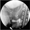
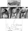
Preoperatively, he was evaluated with simultaneous retrograde and antegrade urethrography to evaluate the exact location and length of the stricture. The images revealed a complete posterior urethral disruption, and the defect length was 4 cm (Fig. 1). Pseudomonas grew in urine cultures and intravenous antibiotics were administrated for a week before the operation. Under general anesthesia, he was placed in a lithotomy position. Buccal mucosal grafts were harvested from both inner cheeks by a plastic and reconstructive surgery team. The inner cheek was infiltrated with 1:200,000 epinephrine and two 5.0×2.5 cm and 5.0×2.0 cm sized grafts were harvested. The grafts were defatted and tubed around a 20 Fr Foley catheter by use of interrupted 4-0 polyglactin sutures (Fig. 2A). The anastomosis of the graft to the distal end of the urethra was performed after resection of fibrotic tissue through a midline perineal incision. To minimize the morbidity of the urethral sphincter, an 18 Fr sound was placed on the proximal urethral end through the suprapubic route, and from the defected segment to the perineum was carefully perforated along with finger palpation of the sound tip (Fig. 2B). After careful dilation up to 24 Fr, a 20 Fr Foley catheter with the tubal buccal mucosa graft was successfully interposed in the defective urethral segment (Figs. 2C-E). All layers were closed after meticulous bleeding control. Suprapubic drainage continued until patency of the urethra could be ensured by postoperative imaging.
The intraluminal Foley catheter was removed 3 weeks after the operation, and while the suprapubic cystostomy was clamped, the patient was instructed to void. He was able to void successfully without urinary incontinence. A postoperative urethrogram showed a good urethral shape with no urinary leaks (Fig. 3A). Although the maximum uroflow rate was weak (13.7 ml/sec), postoperative uroflowmetry showed a normal shape with no residual urine (Fig. 3B). After 8 months he had normal sexual and voiding functions without urinary incontinence.
DISCUSSION
Herein, we reported a successful operation using a tubal buccal mucosa graft without anastomosis of the proximal urethra for managing a long segment posterior urethral defect. This procedure has several advantages. First, injury to perineal structures can be minimized owing to the avoidance of anastomosis of the proximal urethral end. Of course, direct end-to-end anastomosis between the proximal and the distal healthy urethral mucosa is the best method for a successful operation. However, in terms of a complicated long segment posterior urethral defect, extensive dissection is inevitable for approach to the proximal urethral end. Especially in the case of sexually active young patients, preservation of the perineal nerves is of great importance for preserving erectile function. In addition, sphincter injury can be diminished because only staged dilatation using a sound is performed in the urethral sphincter without incision or excision of muscles.
Surgical principles for posterior urethral stricture caused by pelvic bone fracture are excision of the fibrotic scar and a tension-free anastomosis. Complicated cases such as patients with prior failed surgeries or long segment strictures may need extensive surgery, such as a combined abdominal-perineal approach with complete pubectomy for adequate length and a tension-free anastomosis. However, this could be associated with urinary incontinence, impotence, or even failure again. Moreover, our patient was young and had normal erectile function; thus, we believed that aggressive surgery was not appropriate.
Nowadays, the most commonly used graft materials for a refractory urethral stricture are skin, bladder mucosa, and buccal mucosa. El-Sherbiny et al. [4] studied 3 graft materials in experimental dogs and concluded that buccal mucosa was the best graft material among them. They reported that buccal mucosa grafting was associated with the lowest stricture rate of 12%, followed by 37% for bladder mucosa and 62% for skin, and graft shrinkage was less than 10% for buccal mucosa compared with 20 to 40% for skin and bladder mucosa. Buccal mucosa has a thick epithelium rich in elastin that makes it easy to handle and durable [5]. In clinical studies, the overall success rate of buccal mucosa was higher than for skin grafting [6]. Considering the physiology, bladder mucosa grafts may be the best material for contact with urine. However, bladder mucosa has been associated with many complications, including stricture, prolapse, and a granulomatous reaction [7]. The lamina propria of buccal mucosa is thin compared with bladder mucosa and skin, which facilitates inoculation and neovascularization. Moreover, additional abdominal incisions are needed to harvest the bladder mucosa. In terms of graft techniques, onlay grafts usually have better results than tubal grafts [4]. Tubal grafts may fail because of inadequate graft take, because they are not circumferentially surrounded by vascularized tissue, and because of inexperience of the surgeons [1]. However, considering the location and length of the stricture in our case, pubectomy and a combined abdominal-perineal approach would have been required for proper onlay technique. Therefore, we believed that a tubal buccal mucosa graft was the most suitable method for our case.
For the best results with tubal grafts, anastomosis of both ends with healthy urethral tissue is recommended. To minimize morbidity of the urethral sphincter, however, in our case, the tubal buccal mucosa graft was interposed in the urethral defect without anastomosis of the proximal end. We expected that the floating buccal mucosa of the proximal site would be denuded owing to a lack of blood supply. Although long-term follow up is needed, this method could successfully preserve the external sphincter and sexual functions.
In conclusion, tubal buccal mucosa graft without proximal anastomosis could be an alternative method for long posterior urethral defects involving the external sphincter.

 XML Download