

 PDF
PDF ePub
ePub Citation
 Print
Print


INTRODUCTION
Globally, prostate cancer (PCa) is one of the most prevalent forms of malignancy diagnosed in elderly men; moreover, it is the most common cancer and the second most common cause of cancer death in the United States and the United Kingdom [1]. In Korea the incidence of PCa was 26.1 per 100,000 men during 2008 and had increased annually by 13.7% from 1999 to 2008 [2].
Most PCas are diagnosed by transrectal ultrasound (TRUS)-guided prostate needle biopsy, which is indicated in men with an elevated serum prostate-specific antigen (PSA) level, abnormal digital rectal examination (DRE) finding, or hypoechoic lesions in TRUS. However, not all PCas are diagnosed at the initial biopsy. Therefore, in men with a negative initial biopsy result and persistent clinical suspicion of prostate cancer, a repeat biopsy is indicated [3].
Previous studies have shown that PCas diagnosed at repeat biopsy are smaller and are less likely to be high grade as shown by examination of the prostate needle biopsy specimen [4] and are related to better pathological outcomes after radical retropubic prostatectomy (RRP) [5-7]. Furthermore, repeat prostate biopsy patients are more likely to harbor clinically insignificant PCa or indolent cancer [5,8-10]. With regard to the nature of PCa diagnosed at repeat biopsy, controversies remain because some of the literature has reported that the Gleason score (GS), stage, and tumor volume of PCas detected on initial and repeat biopsy are similar [11].
However, there are few published data on the characteristics of PCa detected on repeat biopsy in Korea. In our study, we analyzed the differences in pathological outcomes in the RRP specimens on the basis of whether PCa was detected at the initial or repeat biopsy.
MATERIALS AND METHODS
We retrospectively reviewed the medical records of 287 PCa patients who underwent RRP from January 2005 to December 2010 and did not have a history of prior radiotherapy or hormonal therapy.
We collected preoperative clinical factors including age, serum PSA level, prostate volume (PV) on TRUS, DRE findings, biopsy schema (number of cores taken), clinical stage (2002 American Joint Committee for Cancer tumor-node-metastasis staging system), and number of prior prostate biopsy procedures. In addition, postoperative pathological outcomes such as specimen volume, percentage tumor volume, GS, tumor bilaterality, pathological stage, and presence of positive surgical margin (PSM)/lymphovascular invasion (LVI)/perineural invasion (PNI) were investigated.
A repeat biopsy was performed in patients with an abnormal DRE result, persistently increased or increasing (PSA velocity greater than 0.75 ng/ml/yr) PSA level, low free PSA, or prior atypical small acinar proliferation or high-grade prostatic intraepithelial neoplasia. However, each of us had a different indication for repeat biopsy.
With regard to biopsy schema, TRUS-guided sextant biopsy cores were taken until 2007; after that, 10- to 12-core biopsy was performed. In all repeat biopsy cases, however, a 10- to 12-core biopsy schema was used.
RRP specimens were reviewed by a single experienced pathologist. After the seminal vesicles were removed, fresh specimens were weighed and the fresh weight was regarded as specimen volume (actual prostate weight). Prostate specimens were then entirely embedded and serially sectioned at 3-mm intervals. On all slides involved with tumor, the tumor area was marked and the percentage tumor volume (tumor percentage for the entire prostate) was calculated. Percentage tumor volume was determined by the average of the sum of the results from all slides involved with tumor.
Patients were divided into two groups by the number of biopsies before diagnosis (initial biopsy vs repeat biopsy: at least two biopsies). We then compared preoperative clinical factors and postoperative pathological outcomes between the two groups. The Mann-Whitney U test was used for comparisons of continuous variables and the chi-square test was used for categorical variables. Multivariate logistic and linear regression analyses were used to assess the correlation between repeat biopsy and postoperative pathological outcomes. Statistical analysis was performed with SPSS ver. 15.0 (SPSS Inc., Chicago, IL, USA) and statistical significance was defined as a p<0.05.
RESULTS
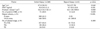
Of the 287 patients, 246 (85.7%) were diagnosed with PCa at the initial biopsy and 41 (14.3%) at the repeat biopsy. In the repeat biopsy group (41 patients), 26 patients (9.1%), 10 patients (3.5%), and 5 patients (1.7%) were diagnosed with PCa at the second, third, and fourth biopsy, respectively. The preoperative characteristics of the 287 patients stratified by the number of prior prostate biopsy procedures are summarized in Table 1. Patients with PCa diagnosed at a repeat biopsy were significantly older (70.0 vs. 67.8, p=0.048) and had a larger PV (42.1 cm3 vs. 35.2 cm3, p=0.009) than did patients with PCa diagnosed at the initial biopsy and had a significantly different biopsy schema (p<0.001). No significant differences were noted between the two groups in serum PSA level, DRE findings, or clinical stage.
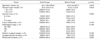
Table 2 presents the univariate analysis of pathological outcomes in patients according to the number of prostate biopsies. There were no significant differences in specimen volume, GS, tumor bilaterality, pathological stage, or status of PSM/LVI/PNI. Regarding GS, the incidence of a GS of 6 or less, a GS of 7, and a GS of 8 or more was 47.5%, 42.5%, and 10.0%, respectively, in the repeat biopsy group compared with 30.2%, 60.4%, and 9.4%, respectively, in the initial biopsy group, reflecting a trend toward low GS disease in the repeat biopsy group. However, this trend did not reach statistical significance (p=0.082). Concerning percentage tumor volume, 72.5% of the repeat biopsy group had a percentage tumor volume <10% compared with 51.6% of the initial biopsy group, and 27.5% of the repeat biopsy group had a percentage tumor volume ≥10% compared with 48.4% of the initial biopsy group. Thus, the repeat biopsy group had a lower (<10%) percentage tumor volume (p=0.016).
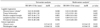
The multivariate analysis (after adjustment for biopsy schema, age, serum PSA, PV, and DRE) showed that repeat biopsy was an independent predictive factor of lower (<10%) percentage tumor volume (p=0.037). However, repeat biopsy was not an independent predictor of GS, tumor bilaterality, pathological stage, PSM, LVI, or PNI (p=0.212, 0.456, 0.459, 0.917, 0.991, and 0.827, respectively) (Table 3).
DISCUSSION
According to recent studies, 20 to 30% of patients are diagnosed with prostate cancer at the initial biopsy [5,6]. Because a negative initial biopsy does not mean that patients have no PCa, repeat biopsy has been a major concern for urologists and patients. Although there is no definitive guideline on repeat biopsy in men with an initial negative biopsy result, a repeat biopsy is usually indicated in men with a persistently increased PSA level, PSA velocity greater than 0.75 ng/ml/yr, low free PSA, abnormal DRE, or previous pathological findings related to an increased risk of cancer, such as atypical small acinar proliferation or high-grade prostatic intraepithelial neoplasia [4-8,12-17].
The published studies on PCa patients who have undergone RRP have reported that 72.6 to 92.5% of PCas are detected at the initial biopsy, and the remainder are diagnosed at a subsequent biopsy [5-8,11]. In our study, of the 287 PCa patients, 246 patients (85.7%) were diagnosed with cancer at the initial biopsy and 41 patients (14.3%) at the repeat biopsy. This result shows that our diagnostic yield of prostate biopsy is comparable with that of previously published studies.
In our study, the repeat biopsy group had a significantly larger PV (p=0.009) than did the initial biopsy group and showed no significant difference in biopsy schema (p<0.001). In many previous studies, PV was significantly higher in the repeat biopsy group [4,5,8,11] and the biopsy schema was not identical during the time period [6-8]. For these reasons, we adjusted for PV and biopsy schema in the multivariate analysis.
There is some controversy about the characteristics of PCa detected at repeat biopsy, although favorable outcomes have prevailed. In 2006 Lopez-Corona et al. [5] studied 1,357 patients treated with RRP. Patients with 2 or greater biopsies had a higher rate of clinical T1c stage cancer and larger prostates than did patients with only 1 biopsy (each p<0.0001). After RRP, patients with 1 biopsy had a lower rate of organ-confined tumors (61% vs. 75%, p<0.0001) and a higher rate of extracapsular extension, seminal vesicle invasion, lymph node metastases, and a GS of 7 or greater than did other patients. Indolent cancer was found in 10% of patients with 1 biopsy and 18% of those with 2 or more biopsies (p=0.018). Those authors concluded that PCa diagnosed after repeat biopsy has a favorable pathological outcome [5]. Resnick et al. [8] also reported that patients undergoing multiple prostate biopsies before RRP are more likely to harbor clinically insignificant prostate cancer than are those who undergo only 1 biopsy before RRP. In Korea, Park et al. [6] found that patients with repeat biopsy had higher rates of clinical T1c disease (79.5% vs. 55.5%, p<0.001), higher rates of pathologically organ-confined disease in the RRP specimen (78.3% vs. 61.3%, p=0.003), lower rates of a GS≥7 (63.7% vs. 73.7%, p=0.029), a lower number of positive cores (2.3 vs. 3.1, p<0.001), and a lower tumor volume in RRP specimens (4.4 ml vs. 7.8 ml, p<0.001) than did patients with an initial biopsy only. As contrasted with the above studies, in the European Prostate Cancer Detection study, Djavan et al. [11] reported no significant differences with respect to GS, percentage Gleason grade 4/5, pathological stage, and tumor volume between the initial and the repeat biopsy group. They concluded that PCa detected on repeat biopsies has similar biological properties and at least identical characteristics as PCa found at the initial biopsy. The results of our study are similar to those of the European Prostate Cancer Detection study. We found that there were no significant differences in clinical stage between the initial and the repeat biopsy group. Furthermore, in the RRP specimens, the GS, tumor bilaterality, pathological stage, and PSM/LVI/PNI status of the repeat biopsy group were not significantly different from those of the initial biopsy group (p=0.212, 0.456, 0.459, 0.917, 0.991, and 0.827, respectively). The only difference was that percentage tumor volume differed significantly; the repeat biopsy group had a lower (<10%) percentage tumor volume (p=0.016). The reason percentage tumor volume was dichotomized at 10% was that our data for percentage tumor volume showed a statistically significant difference at this level and this cutoff point has previously been shown to be one of the factors determining clinically insignificant PCa. As in our study, Resnick et al. [8] reported that an increasing number of prior prostate biopsies was directly associated with the risk of low-volume disease. Indeed, 18.8% of the initial biopsy group was found to have an estimated tumor volume (percent tumor volume in our study) <2% compared with 35.1% of the group with 3 or more biopsies. In addition, 42.4% of the initial biopsy group was found to have estimated tumor volume >10% compared with 29.3% of the group with 3 or more biopsies (p<0.01).
Unfortunately, we as well as Resnick et al. [8] could not address tumor volume itself because we retrospectively reviewed the medical records, in particular, pathological reports that had already been described. Furthermore, we could not investigate unusual sites (such as the transitional zone, anterior fibromuscular zone, far lateral peripheral zone, and adjacent site of seminal vesicle) of PCa and did not estimate biochemical-free survival for the same reason. Another limitation of the present study was that there is a likelihood of selection bias because our study included only the patients who underwent RRP instead of all patients with biopsy-confirmed PCa. Therefore, localized PCas with low PSA, low grade, and low stage were mainly included. Finally, in our study, the biopsy schema and indication for repeat biopsy were not standardized during the 6-year period studied.
In conclusion, although the above-mentioned limitations existed in our study, we found that postoperative pathological outcomes of PCas detected at repeat biopsy were not significantly different from those of PCas detected at the initial biopsy except that the percentage tumor volume was lower (<10%) in PCas diagnosed at repeat biopsy.
CONCLUSIONS
Other than in a few studies, most previous studies demonstrated that patients diagnosed with PCas at a repeat biopsy had more favorable pathological outcomes than did patients with PCas diagnosed at the initial biopsy. However, we found no significant differences in specimen volume, GS, tumor bilaterality, pathological stage, or PSM/LVI/PNI status between the initial and repeat biopsy groups, except that the percentage tumor volume was lower (<10%) in the patients diagnosed with prostate cancer at a repeat biopsy.
Further investigation including the evaluation of tumor volume itself and an analysis of the survival benefit of percentage tumor volume should be performed.
 XML Download