

 PDF
PDF ePub
ePub Citation
 Print
Print


INTRODUCTION
Global estimates suggest that one in every three males worldwide is circumcised [1]. According to advocates of the procedure, circumcision may also provide health benefits to males, the most prominent being the prevention of human immunodeficiency virus infection [2]. Many studies have shown that male circumcision also protects against urinary tract infections in infants and children [3].
Urinary tract infection is a common bacterial infection in childhood and causes significant morbidity [4,5]. Among the significant complications are concomitant bacteremia, acute failure, and progressive renal damage [6]. Many reports have linked the risk of developing urinary tract infection with the circumcision status of the child [5]. The uncircumcised state is associated with a higher incidence of urinary tract infection, especially in high-risk populations like infants and children with congenital anomalies [4]. However, there is no universal acceptance that circumcision prevents the occurrence of urinary tract infection [7].
In this study, we evaluated the effect of the preputial type on bacterial colonization and wound healing in boys undergoing routine circumcision in accordance with religious belief [8].
MATERIALS AND METHODS
The study consisted of 78 boys admitted to our clinic for circumcision consecutively between 2009 and 2011. Informed consent was obtained from the patients at the beginning of the study. With the subject relaxed and supine, the prepuce was gently retracted without traumatic force and the degree of preputial retractability was determined. Preputial status was classified into five types on the basis of preputial retractability as follows: type 1: no retraction of prepuce at all, type 2: external urethral meatus exposure only, type 3: glans exposure halfway to the sulcus of the corona, type 4: glans exposure to above the corona at the site of preputial adhesion, and type 5: easy exposure of the whole glans [8]. The indication for surgery for all patients was religious belief. Before the surgery, preputial cultures were taken after the foreskin was retracted carefully in order to avoid external contamination. These cultures were taken from each child by the same investigator. No external cleansing was performed before the sampling. One sterile culture swab was swept circumferentially once around the surface of the glans starting just proximal to the urethral meatus. Control swabs from the same region of the same patients were taken and inoculated after 3 weeks following circumcision. Thus, the same patients formed the control group. None of the children received antibiotics during this period.
The circumcision procedure of all the patients was performed under general anaesthesia. The circumcision procedure was performed with total removal of the foreskin in all patients. Briefly, after full retraction of the preputium and exposure of the whole glans, a circular incision was performed initially at the mucosal surface of the preputium about 5 mm proximal to the glans. Then the preputium was retracted and another circular incision was performed at the level of the coronal sulcus in the outer skin. The subcutaneous fatty tissue was trimmed and the foreskin marked by the two incisions was removed completely.
Preputial cultures were taken with sterile cotton swabs before cleansing with 10% polyvidone-iodine solution. These were transferred to the laboratory and suspended in 1 ml of 0.9% saline solution in a sterile tube. The solution was then diluted 100 times by transferring 0.1 ml of the solution to another tube filled with 9.9 ml of 0.9% saline solution. An amount of 10 µl of the final solution was inoculated on to 5% sheep blood agar and Eosin-methylene blue agar. The plates were incubated at 37℃ for 24 to 48 hours, after which the different bacterial colonies were counted. Cultured bacteria were identified according to colony morphology, gram staining, and biochemical characteristics by use of standard techniques. Clinically significant bacterial colonization was arbitrarily accepted as the presence of the growth of ≥100,000 colony-forming units per milliliter (cfu/ml) before cleansing with 10% polyvidone-iodine solution. Post circumcision cultures were taken from the suture line 3 weeks after the operation.
To assess whether preputial type affected wound healing, patient wounds were checked for local swelling, local hyperemia, local temperature increase, and local fluid collection on days 1 and 7 after the operation. We evaluated the local swelling on a scale from 0 to 1. The same evaluation scale was used for local hyperemia, local temperature increase, and local fluid collection to determine possible statistical differences between the types.
Data analyses were carried out on a personal computer with the use of SPSS ver. 15.0 (SPSS Inc., Chicago, IL, USA). The statistical significance level was set at 0.05.
RESULTS
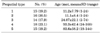
The mean age of the children was 46.3 months (range, 1 to 168 months). Classifications by preputial type and the mean age of patients are shown in Table 1. There was a significant difference among preputial types in terms of mean age (p<0.005). As one variable increased, the other also increased.
A total of 28.2% of the uncircumcised boys had no growth in their preputial culture, 56.4% had pathogenic bacterial colonization, and 15.4% had skin commensals.
Table 2 shows the ratios of uropathogenic microorganisms found in the preputial culture. This ratio was 71.8% in the pre-circumcision culture. The distribution of the ratios of microorganisms according to preputial types is given in Table 2. There was a significant difference in terms of growth (p<0.001).
Of the patients with a pathogenic bacterial colonization in their pre-circumcision antibiogram, three patients of type 1, two patients of type 2, one patient of type 3, one patient of type 4, and one patient of type 5 had skin flora in their third week control culture antibiograms. Patients with a pathogenic bacterial colonization and those with skin commensals were the same individuals. Following circumcision, none of the patients had pathogenic microorganisms in their control culture antibiograms.
When we evaluated the wounds of the patients 1 day after the operation, all had local swelling and hyperemia; these findings were especially noticeable in type 1, 2, and 3 patients. Seven days after the operation, 71.2% of the type 1 patients had swelling and 70.1% had hyperemia, 68.2% of the type 2 patients had swelling and 69.1% had hyperemia, and 45.6% of the type 3 patients had swelling and 35.1% had hyperemia. No swelling was encountered in type 4 or 5 patients. Seven days after the operation, there was a significant difference in terms of wound healing. This difference was observed in all types (p<0.05) except type 1 and type 2 (p>0.05). Patients did not have local fluid collection on either postoperative day 1 or day 7. While taking the post-circumcision culture, it was noticed that all patients had complete recovery, and no complications were encountered.
DISCUSSION
Circumcision is an operation with a long history [9]. It may provide health benefits to males, the most prominent being prevention of human immunodeficiency virus infection. It was recently shown in randomized controlled trials that adult circumcision reduces the risk of acquiring human immunodeficiency virus infection [2]. Many studies have shown that male circumcision also protects against urinary tract infections in infants and children [3]. Urinary tract infections are mostly accepted as arising by the ascending route, and the agents mostly come from the periurethral region [10]. Colonization in the preputial sac may create a reservoir from which the organisms ascend, resulting in bacteriuria and urinary tract infection and inability to retract the preputium. Phimosis promotes this migration by providing an optimum environment for growth of possible uropathogenic bacteria [11]. Many studies designed to evaluate the periurethral flora in uncircumcised boys from birth to 16 years have revealed massive bacterial colonization during early infancy, mainly with Escherichia coli [6]. Similarly, Wiswell et al. reported that the presence of the foreskin was associated with a higher rate of colonization with gram (-) pathogens during the first 6 months of life [11].
Kayaba et al. [8] evaluated 603 Japanese boys between the ages of 0 and 15 years. They found that the degree of preputial separation increased with age. Before age 6 months, the incidence of types 1 and 2 prepuce was 47.1% and 21.5% respectively. Thereafter, type 3 prepuce was most common until preschool age. Of the 11- to 15-year-old subjects, 62.9% had type 5 prepuce. Our study revealed a decreased frequency of unretractable prepuce in the older age group.
The relationship between circumcision and urinary tract infection was initially described by Ginsberg and McCracken in 1982 [7]. A meta-analysis of nine of the major studies demonstrated a 12-fold increase in the risk of urinary tract infection in uncircumcised boys, and that between 0.9% and 4.2% of all uncircumcised boys would have symptomatic urinary tract infection in the first year of life [12]. In the study by Cascio et al. [4], a pure growth of uropathogens was isolated in 37% of uncircumcised boys with vesicoureteral reflux who were receiving antibiotic prophylaxis and in 28% of boys who had undergone circumcision. In another study, the authors isolated pathogenic bacteria in the periurethral region of 64% of patients before circumcision, and this rate decreased to 10% after circumcision [1]. In our study, there was a 71.8% growth rate in pre-circumcision patients and a 10.25% growth rate in the post-circumcision group; however, none of these patients had pathogenic microorganism growth. Staphylococcus aureus is known as the most frequently cultured bacteria after circumcision, and circumcision status probably does not affect the colonization of these bacteria, as the results of our study also confirmed [13]. Wijesinha et al. [14] showed how the skin flora changed 3 weeks after circumcision from uropathogens to skin commensals. We found the same result in our study. In some of the pre-circumcision patients who had pathogenic microorganisms in their culture antibiogram, 3 weeks after circumcision, the only organisms cultured from the periurethral region were skin commensals. Tokgoz et al. [6] analyzed the preputial flora in preschool and primary school boys and found that significant colonization with possible uropathogens still persisted in this age group in 100% of patients with phimosis and 48.1% of cases without phimosis. Hallett et al. [10] showed no difference in colony counts between boys with phimosis and those without phimosis. Our study revealed a growth of 100% in type 1, 93.8% in type 2, 71.4% in type 3, 44.4% in type 4, and 53.6% in type 5. There was a significant difference between the types in terms of growth (p<0.001).
The most common microorganisms isolated in children with uncomplicated urinary tract infection are of the Enterobacteriaceae family, namely E. coli, Klebsiella, and Proteus species [6]. Glennon et al. [5] found a 22% rate of Proteus mirabilis colonization in uncircumcised boys. We isolated this bacterium in 6.6% of our cases. In their analysis of periurethral bacterial flora before circumcision, Wijesinha et al. [14] isolated Proteus, Coliforms, and Enterococcus species in 52% of boys; we documented these bacteria in 52.6% of our cases. Tokgoz et al. [6] demonstrated significant preputial bacterial colonization. They found E. coli in 3.1% of the cases, Klebsiella species in 18.8%, coagulase-negative Staphylococcus species in 12.5% and Enterococcus species in 43.8%. The most common microorganism in our study was also Enterococcus, in 33% of the cases; the others were E. coli in 12.8% of cases, Proteus species in 6.6%, Klebsiella species in 2.6%, and coagulase-negative Staphylococcus species in 15.4%.
There is no account of a study on wound healing according to preputial types in the literature. In our study, when we evaluated the wounds on postoperative day 1, all patients had local swelling and hyperemia, but type 1, 2, and 3 patients in particular had more noticeable swelling and hyperemia. However, 7 days after the operation, 71.2% of type 1 patients had swelling and 70.1% had hyperemia, 68.2% of type 2 patients had swelling and 69.1% had hyperemia, and 45.6% of type 3 patients had swelling and 35.1% had hyperemia. No swelling was observed in type 4 and 5 patients. We believe that late healing in type 1, 2, and 3 patients results from preputial adhesiveness and bacterial colonization, and parents should be informed of this.
CONCLUSIONS
There is a significant relationship between preputial type and bacterial colonization. However, as patients get older, bacterial colonization can continue even though the preputial type changes. In circumcision, the removal of the whole preputium could eliminate the site for bacterial growth and subsequent risk of infection. Preputial type also affects post-circumcision wound healing. Especially in countries like ours where circumcision is common and obligatory, it should be considered that wound healing takes longer in patients with preputium of type 1, type 2, and type 3, and patients should be informed of this. In our opinion, it is due to preputial adhesiveness and bacterial colonization.

 XML Download