

 PDF
PDF ePub
ePub Citation
 Print
Print


INTRODUCTION
Expectant therapy, extracorporeal shock wave lithotripsy (ESWL), and ureteroscopic removal of stones (URS) could be considered the first-line therapy for ureteral stones. In recent years, although ESWL has been universally adopted for the treatment of ureteral stones, URS remains the choice for initial therapy [1,2]. Reductions in complication rates have been achieved in the past 2 decades as the result of technological advances and increased clinical utilization, and URS is now considered equivalent to ESWL [3,4].
URS is accepted as the treatment of choice for lower ureteral stones, in which stones may be cleared intraoperatively in 80 to 100% of cases, often with 100% stone-free rates by the second day [5]. URS for mid-ureteral or upper ureteral stones provides a high stone clearance rate of more than 90% [6].
With reduced risk of adverse events, urolithiasis has been migrating away from the in-patient setting since the 1990s. In most medical centers in the United States, stone procedures are now performed in the ambulatory setting at hospital outpatient facilities with ESWL and URS as the predominant treatment modalities [7,8].
Despite improved stone-free rates and reduced complication rates through the evolution of the surgical instruments used, postoperative complication rates remain-moderate. The incidence of immediate, unplanned admission owing to post-URS complications ranges from 1.5 to 14.3% in Western countries [9,10]. Postoperative pain is the predominant complication.
Until now, most urologists have not studied acute postoperative pain, which the patients perceive as a serious problem and which accompanies most postoperative complications. Considering that most URS procedures in many countries are performed with the use of a rigid ureteroscope, the incidence of acute postoperative pain and postoperative complications could be higher than in other countries that use a flexible ureteroscope [9].
To better define acute postoperative pain and to decrease the risks of unscheduled admission after URS, we performed a retrospective analysis of the URS experience at our institution to determine the incidence of acute postoperative pain following URS and to identify potential risk factors associated with it. This study aimed to help clinicians implement the most appropriate pain control strategy after URS to reduce unscheduled visits or admissions post-URS.
MATERIALS AND METHODS
This study was part of a prospective longitudinal observational study to investigate acute postoperative pain. From June 2008 to December 2010, URS was performed by a single urologist on 143 patients. Patient preparation, the URS procedure, postoperative care, discharge, and follow-up were done according to our routine URS protocol.
Ureteral stones situated below the lower margin of the sacroiliac joint illustrated radiologically were defined as lower ureteral stones. Upper ureteral stones were defined as stones located between the renal pelvis and the top edge of the sacrum. The size of a ureteral stone was measured by its maximum diameter through simple abdominal radiography, excretory urography, and non-enhanced computed tomography (CT).
Patients were admitted 1 day preoperatively. Prophylactic and postoperative intravenous broad-spectrum antibiotics were routinely administered. URS was performed under general or spinal anesthesia. A 8.5 Fr. rigid ureteroscope (Richard Wolf Medical Instruments Co., Vernon Hills, IL, USA) was used and lithotripsy was performed with a pneumatic lithoclast (Richard Wolf Medical Instruments Co.). Foreign body forceps or a stone basket (COOKMedical, Bloomington, IN, USA) was selectively used. Ureteral orifice dilation was not performed routinely, and if needed, a facial dilator was used. A double pig-tail catheter was inserted at the surgeon's discretion but was placed routinely in cases of a solitary kidney, bilateral procedures, renal insufficiency, and ureteral injury such as perforation.
Assessment of treatment outcome was based on the stone-free rate and the incidence of intraoperative complications. According to our protocol, the visual analogue pain scale (VAS) score (normal range, 0 to 10) was checked with each patient on the first postoperative day and patients were discharged in the afternoon of the first postoperative day. Provided there were no postoperative complications, patients were followed up on the seventh postoperative day and the double pig-tail catheter was removed under local anesthesia on the same day. Patients were advised to visit the emergency unit at any time in the case of postoperative complications, such as pain, clot retention, fever, and voiding difficulties.
After reviewing the records of 143 patients who were treated according to the URS protocol, 8 patients were excluded owing to intraoperative complications. Those patients were regarded as having failed URS despite stone clearance. The remaining 135 patients were divided into two groups according to the absence or presence of acute postoperative pain: group 1, with acute postoperative pain, and group 2, without acute postoperative pain. Acute postoperative pain was defined as a VAS score greater than 4, which suggests moderate to severe pain. Patients with acute postoperative pain were analyzed on the basis of serial postoperative complications and pain control medication. Factors such as age, body mass index, sex, urolithiasis history, hypertension, diabetes mellitus, history of urinary tract infection (UTI) (e.g., acute pyelonephritis, cystitis, acute prostatitis identified through diagnoses or medication within 3 months), presence of psychiatric illness (e.g., depression, anxiety, and indexes that assess psychiatric distress), pyuria, level and size of stone, location and laterality of stone, symptom duration, unilateral or bilateral procedure, solitary kidney, use of lithotripsy, ureteral dilatation, use of a basket or forceps, spinal or general anesthesia, operation time, and placement of a ureteral stent were analyzed to identify potential risk factors that-could predict acute postoperative pain.
In the statistical analysis, Mann-Whitney U tests and Fisher's exact tests were used for univariate analysis of the significance between variables by use of SPSS ver. 13.0 (SPSS Inc., Chicago, IL, USA). Factors were selected for multivariate analysis if the univariate analysis showed a p-value of less than 0.05. Logistic regression models were-applied as appropriate. Differences were considered to be statistically significant when the p-value was less than 0.05 (p<0.05).
RESULTS
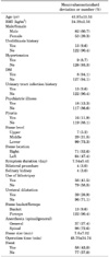
Patient characteristics are summarized in Table 1. The average age of the 135 patients (82 male and 53 female patients) was 43.97±13.53 years old. The overall success rate of URS at our institution was 95.5%. Failure of URS was mainly due to intraoperative complications (Table 2). The overall incidences of acute postoperative pain and postoperative complications were 14.6% and 9.6%, respectively. All patients who experienced postoperative complications also experienced acute postoperative pain.
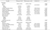
The results of the comparative analysis between the two groups were as follows: age, psychiatric illness, history of UTI, ureteral dilation, use of a stone basket, stone size, and operation time were shown to be potential risk factors in the univariate analysis (p<0.05). In the multivariate analysis, age (p=0.048), psychiatric illness (p=0.007), history of UTI (p=0.002), and ureteral dilation (p>0.001) remained significantly associated with acute postoperative pain.
The median age of group 1 was 38.48 years, compared with 44.98 years in group 2 (p=0.048). Eight patients (38.1%) had a psychiatric illness in group 1, compared with 10 patients (8.8%) in group 2. Eight patients (38.1%) had a history of UTI in group 1, compared with 5 patients (4.4%) in group 2 (p=0.002). Fifteen patients (71.4%) underwent ureteral dilatation in group 1, compared with 24 patients (21.1%) in group 2 (p>0.001). Five patients (23.8%) required a stone basket during surgery in group 1, compared with 8 patients (7.0%) in group 2 (p=0.538). The median stone size was 9.10 mm in group 1 and larger than 7.39 mm in group 2 (p=0.191). The median operation time was 54.52 minutes in group 1, compared with 47.63 minutes in group 2 (p=0.693) (Table 3).
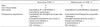
Among the 21 patients with acute postoperative pain, 14 patients complained of moderate acute postoperative pain (VAS, 4 to 6), and 7 patients complained of severe acute postoperative pain (VAS, 7 to 10). Their postoperative course and features are described in Table 4. Ten patients with moderate acute postoperative pain underwent placement of a ureteral stent. On the other hand, no patient with severe acute postoperative pain underwent placement of a ureteral stent (p=0.004). Four patients with severe acute postoperative pain required a stone basket intraoperatively, which was one more than in those with moderate acute postoperative pain. The pain of 11 patients with moderate acute postoperative pain was well controlled with intravenous or intramuscular injection of nonsteroidal anti-inflammatory drugs (NSAIDs), whereas 4 patients with severe acute postoperative pain did not tolerate NSAIDs and needed opioid agents.
DISCUSSION
Advancements have been made in the treatment of ureteral stones, which constitute the majority of urologic conditions. In the past, the treatment of ureteral stones depended mainly on invasive procedures. In recent years, noninvasive procedures, such as ESWL and endourologic surgery, have been made available. The ureteroscope was introduced by Goodman [1] and Lyon et al. [2] in the 1970s and has been developed into much smaller, flexible, pneumatic, and laser lithotripsy instruments after URS with a rigid ureteroscope was reported by Pérez-Castro Ellendt and Martinez-Piñerio in 1982 [11]. For lower ureteral stones, expectant therapy, ESWL, URS, ureteral stenting, and open surgery are available therapeutic methods according to the size of the ureteral stone. ESWL and URS are being administered as first-line interventions in most cases that require active treatment [12,13]. Despite the introduction of ESWL, URS has played a key role in the treatment of ureteral stones. It is being widely used as first-line treatment in the removal of lower ureteral stones. It is necessary to fully understand the anatomical structure of the upper ureter and to master the pathogenesis and management of its complications to increase the success rate of URS. Because URS is an endoscopic procedure, technique, knowledge of the equipment, and operating skills are fundamental to reducing complications. Accurate location of ureteral stones and ureter conditions must be elucidated by excretory urography and ultrasonography before performing URS [14].
The success rate of URS depends on the size of the ureteral stone, its location in the ureter, the utility of surgical instruments including the ureteroscope, and surgical technique. Early reports on the success rate of URS varied from 57 to 97%. The success rate by ureteral stone location was reported to range from 22 to 60% in the upper ureter, from 36 to 83% in the mid-ureter, and from 84 to 99% in the lower ureter [15]. This study yielded a comparatively high success rate of 97%. General or spinal anesthesia is needed in URS to prevent ureteral damage secondary to patient movement intraoperatively and to facilitate stone removal by relaxing the ureteral muscle and urogenital diaphragm [16].
Lee et al. [17] reported the causes of URS failure to include failure of insertion of the ureteroscope into the ureter, migration of ureteral stones toward the upper end, failure of ureteral dilatation, and ureteral stone crushing. Failure of ureteroscope insertion into the ureter is a major cause of failure that occurs when a ureteral orifice is too narrow to facilitate ureteroscopic entry and when the angle is too large between the lower urinary tract, including the ureteral orifice, and the ureteroscope indwelled in a bladder. Excessive extension of a ureter and entry into aureteroscope increases the risk of ureteral damage. Green and Lytton [18] reported the causes of URS failure to include ureteral stricture, bleeding, migration of ureteral stones toward the upper end, and a pseudo-ureteral channel of the bladder.
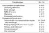
URS complications can be divided into early and late complications. Early complications include ureteral perforation during surgery, damage to the ureteral mucosa, theformation of a false passage within the ureteral wall, hemorrhage, ureteral tract infection, high fever, sepsis, and temporary ileus. Late complications include ureteral stricture and vesicoureteral reflux [15,17,19]. In a publication that included 1,696 cases of URS, Huffman [20] reported that about 9% of patients experienced complications after surgery, and of those, 1.6% needed surgical intervention. There were 25 cases (14.7%) of intraoperative complications, such as ureteral perforation, false passage, and difficult access, and 29 cases (17.1%) of postoperative complications, such as pain, UTI, urinary retention, and hematuria in this study. However, surgical intervention was not required following URS in this study.
There had been scant research on the risk factors of pain after surgery as an early complication. The mechanism of acute pain after URS remains unclear. Acute postoperative pain is mainly associated with the operation itself [9]. Pain due to urolithiasis is usually caused by acute distention of the renal capsule, generally from inflammation or obstruction, or results from acute distention of the ureter and by hyperperistalsis and spasm of the ureteral smooth muscle as it attempts to relieve the obstruction [21]. During URS, irrigation solution may lead to hydroureter and hydronephrosis, which can be exacerbated by a long operation time. In addition, it appears that stone size, ureteral dilatation, and use of a stone basket are risk factors for early postoperative pain. It is suspected that operation time directly or indirectly causes pain through aggravated ureteral edema, hydronephrosis, and hydroureter damage to the ureteral mucosa.
Cheung et al. [9] asserted that pain and complications increased when surgery time is greater than 60 minutes, and that pain and complications were increased in patients who received ureteral stents in 329 cases of URS conducted on outpatients. In another study, el-Faqih et al. [22] reported that dysuria and pain were associated with ureteral stenting in 79% and 29% of patients, respectively. This study concluded that long surgery time was associated with early postoperative pain, but ureteral stenting was not.
There has been intense research activity to determine whether placement of ureteral stents is required. Hosking et al. [23] asserted that placement of ureteral stents is unnecessary in the management of mid, upper, and lower ureteral stones if there are no complications during and after URS [24]. In this study, placement of ureteral stents was randomly performed at the surgeon's discretion, except in cases of the indications discussed above. Stent placement was not a risk factor for acute postoperative pain but it is strongly assumed that placement of stents could prevent the extreme pain of VAS scores greater than 7. Among 21 patients with acute postoperative pain, no stented patient reported VAS scores of greater than 7.
In psychiatric, anesthetic, and geriatric studies about postoperative pain, other external operation factors considered have included catastrophizing, mood disorders, and age. Catastrophizing is a predictor of postoperative pain intensity according to Papaioannou et al. [25]. Ip et al. [26] reported that preexisting pain, age, anxiety, and type of surgery are significant predictive factors for postoperative pain. Pain in the elderly tends to be constant, be of moderate to severe intensity, last for several years, and be multifocal and multi-factorial in origin. Age was commonly found to have negative correlation with postoperative pain intensity in the geriatric population [27]. In this study, age and psychiatric illness were risk factors for acute postoperative pain. Tan et al. [7] reported the risk factors of immediate unplanned admission, and psychiatric illness was among them. Anxiety has been advocated as a factor in lowering the pain threshold, thus facilitating overestimation of pain [28].
This study has several limitations. Although the study was designed as a prospective study initially, longitudinal follow-up after the first follow-up turned out to be impossible. The main reason was the nature of ureteral stones. After complete resolution of the stones and the associated symptoms, the patients did not feel obligated to be followed up. An additional limitation is that the nonparametric size of the patient population yielded limited analysis.
However, this study identified that acute postoperative pain is related to the satisfaction of the patients. This study revealed that age, psychiatric illness, UTI history, ureteral dilatation, stone size, use of a stone basket, and operation time were associated with acute postoperative pain. However, further research on stone size and the cutoff value of surgery time is needed from a large patient cohort.
CONCLUSIONS
The results of this study suggest that URS is an effective treatment for ureteral stones but can induce pain in a considerable number of patients postoperatively. The risk factors associated with early pain after surgery are age, psychiatric illness, history of UTI, ureteral dilatation, size of the stone, use of a basket, and operation time. Our results suggest that more active pain control should be considered for the patient who has these risk factors.
 XML Download