

 PDF
PDF ePub
ePub Citation
 Print
Print


INTRODUCTION
Bladder cancer is the most common malignancy of the urinary tract [1,2]. Approximately 75 to 85% of patients with bladder cancer are diagnosed as having as non-muscle-invasive bladder cancer (NMIBC). In NMIBC, approximately 70% of patients present as pTa, 20% as pT1, and 10% as carcinoma in situ (CIS) [3]. Generally, 30 to 80% of NMIBC will recur and 1 to 45% of cases will progress to muscle-invasive bladder cancer within 5 years [4-6]. Of NMIBC, T1 transitional cell carcinoma (TCC) of the bladder with lamina propria invasion is known to have different degrees of aggressiveness, with a progression rate varying from 12 to 54% [7,8]. For T1 TCC of the bladder, it is important to select those patients who have a favorable prognosis and those who are at high risk for progression. Thus, many clinical (age, gender, multifocality, tumor size) and pathologic (concomitant CIS, tumor grade, architecture) factors for recurrence and progression have been extensively studied over the years to predict prognosis and guide management decisions [3,6].
Younes et al. [9] were the first to use the muscularis mucosa (MM) in transurethrally resected biopsy specimens to substage T1 TCC of the bladder. The MM is a scattered muscular fiber that is formed as a continuous or interrupted layer running along large blood vessels in the lamina propria between the epithelium and the detrusor muscle. Since that time, several studies have been conducted to identify whether the depth of lamina propria invasion is a valuable prognostic factor with the use of the MM as a landmark for substaging. Several reports have demonstrated a worse prognosis for T1 tumors with deep lamina propria invasion, with progression rates ranging from 29 to 55% [8-12]. However, Platz et al. [13] stated that substaging of early invasive bladder cancer is technically difficult and does not have additional prognostic value with respect to survival.
In this study, we retrospectively reviewed our single-center experience in patients with primary T1 TCC of the bladder and evaluated the feasibility and the prognostic significance of the depth of lamina propria invasion on recurrence and progression.
MATERIALS AND METHODS
Between January 1999 and December 2009, 651 patients underwent transurethral resection (TUR) procedures for bladder tumors at our institution. Among these patients, 184 patients were diagnosed as having primary T1 TCC of the bladder and were followed up for at least 12 months. Of 184 patients, 1 patient who had an immediate radical cystectomy was excluded. Thus, the medical records of the remaining 183 patients (149 men and 34 women) were retrospectively reviewed. The mean patient age was 63.5 years (range, 27 to 93 years). None of the patients included in this study had an upper urinary tract malignancy at diagnosis. After TUR, all patients routinely received intravesical bacillus Calmette-Guerin (BCG) instillation once per week for 6 weeks except for 3 patients. The first patient received Mitomycin-C owing to a previous operation history of kidney transplantation, the second patient had current active pulmonary tuberculosis, and the third patient refused BCG therapy.
All TUR specimens from the routine histologic stains (hematoxylin-eosin) were reviewed again by one genitourinary pathologist. Substaging was defined according to the depth of lamina propria invasion by using the Younes et al. [9] classification as follows: T1a, invasion of the lamina propria superficial to the level of the MM; T1b, invasion to the level of the MM; T1c, invasion through the level of the MM but superficial to the muscularis propria (Fig. 1). When the MM could not be found and the vascular plexus was identified, it served as a marker of MM level [8,14]. Tumors were staged by using the 2002 tumor-node-metastasis staging system [15] and were graded as high or low grade according to the 2004 World Health Organization/International Society of Urological Pathology grading system [16]. The microscopic tumor architecture was divided into papillary and solid. A solid growth pattern refers to a tumor that is composed of diffusely growing sheets of tumor cells without fibrovascular core formation. To classify a tumor as solid, at least 10% of the components need to be judged as solid [17].
The median follow-up period was 36 months (mean, 43.5 months; range, 12 to 146 months). Patients were followed until death or last visit and the follow-up included urinalysis, cystoscopy, and urine cytology every 3 months for the first 2 years, every 6 months for the subsequent 2 years, and then annually thereafter. Abdominopelvic computed tomography was performed annually during the follow-up or when clinically indicated.
Clinical outcome was evaluated in terms of tumor recurrence or progression. Recurrence was defined as the histological detection of TCC of the bladder through TUR or bladder biopsy after the first TUR. Progression was defined as the development of muscle-invasive disease or distant metastasis. The diagnosis of an advanced tumor of the upper urinary tract was not considered as progression [8,17].
Univariate and multivariate analyses were performed by using the log-rank test or the Cox's proportional hazards regression model, respectively. The association between recurrence or progression and clinicopathological factors and the relationship between substaging and other prognostic parameters were examined by using the chi-square test. The prognostic parameters assessed were clinical (gender, multifocality, and tumor size) and pathologic (concomitant CIS, tumor grade, microscopic tumor architecture, and substaging) factors. All statistical analyses were performed by using SPSS ver. 13.0 (SPSS Inc., Chicago, IL, USA). Values of p<0.05 were considered to be statistically significant in all of the analyses.
RESULTS
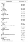
The clinicopathological characteristics of the 183 patients with primary T1 TCC of the bladder are summarized in Table 1. The number of tumors was 2 or more in 87 cases (47.5%), the mean number of tumors was 2.1 (range, 1 to 9), and tumor size was 3 cm or more in 85 cases (46.4%). Concomitant CIS was present with the main tumor in 64 cases (35.0%), a high grade of tumor was found in 171 cases (93.4%), and the microscopic tumor architecture was of the papillary growth pattern in 175 cases (95.6%). Substaging was possible in all 183 cases. Substaging was as follows: T1a in 119 (65.0%), T1b in 57 (31.1%), and T1c in 7 (3.9%). Eight patients (4.4%) underwent radical cystectomy after TUR owing to frequent recurrence or progression.
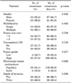
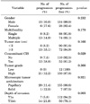
To evaluate the risks of recurrence and progression, T1b and T1c tumors were pooled together because only 7 cases had T1c tumors. The recurrence rate was 32.8% (39/119) for T1a and 40.6% (22/57 for T1b and 4/7 for T1c) for deep lamina propria-invasive tumors including T1b and T1c (T1b/c) during the follow-up period, which was not significantly different (p=0.338) (Table 2). The overall recurrence rate was 35.5% (65/183). The average period until the first recurrence was 16.3 months (range, 1 to 77 months) and the mean number of recurrences was 1.5 times (range, 1 to 4 times). The progression rate was significantly higher in patients with deep lamina propria invasion (p=0.003): 5.8% (7/119) in T1a and 21.9% (14/57 for T1b and 0/7 for T1c) in T1b/c (Table 3). The overall progression rate was 11.5% (21/183). The average period until the first progression was 18.3 months (range, 4 to 69 months). Recurrence and progression rates were not significantly different between low- and high-grade lesions in T1a tumors (p=0.190 and p=0.361, respectively), and those variables could not be compared between low- and high-grade T1b/c tumors because there were no low-grade lesions in T1b/c tumors.
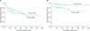
The cancer-specific mortality rate was also significantly higher in patients with deep lamina propria invasion (p=0.036): 4.2% (5/119) in T1a and 14.0% (8/64) in T1b/c. Eight patients died of unrelated causes, giving an overall mortality rate of 11.5% (21/183). Tables 2 and 3 summarize the analysis of the clinicopathological factors influencing recurrence and progression. According to the Kaplan-Meier analysis, the progression-free interval was significantly shorter in patients with T1b/c tumors than in those with T1a tumors (p=0.001). The recurrence-free interval, in contrast, was similar for both groups (p=0.235). Curves for progression-free and recurrence-free estimates are plotted in Fig. 2.
In the univariate analysis of prognostic factors influencing recurrence, microscopic tumor architecture was the only significant prognostic factor for recurrence (p=0.029). However, it was not statistically significant in the multivariate analysis (p=0.075). Also, the depth of lamina propria invasion was not a statistically significant prognostic factor influencing recurrence in either the univariate or the multivariate analysis (p=0.235, p=0.797, respectively) (Table 4).
In the univariate and multivariate analyses concerning progression, depth of lamina propria invasion was a statistically significant prognostic factor (p=0.001 and p=0.006, respectively) and the presence of concomitant CIS was also an independent prognostic factor (p=0.026 and p=0.047, respectively) (Table 5).
The correlation between T1 substaging and other prognostic parameters was evaluated (Table 6). Multifocality, tumor size, and presence of concomitant CIS were similar in the two groups. However, high tumor grade was significantly more frequent in the T1b/c group (p=0.009) and a microscopic solid growth pattern was also significantly more frequent in the T1b/c group (p=0.003).
DISCUSSION
The presence of MM in the lamina propria was first reported by Dixon and Gosling [18] from the biopsy specimens of 25 patients and the radical cystectomy specimens of 4 patients in 1983. The use of MM to substage T1 TCC of the bladder was first described by Younes et al. [9]. Since then, several studies about the MM have been conducted to identify prognostic significance. The results of the present study show that the depth of lamina propria invasion is a significant predictor of progression in patients with primary T1 TCC of the bladder, with a 21.9% progression rate for T1 tumors deeply invading the lamina propria (T1b/c) as opposed to a 5.8% progression rate for T1 tumors without deep lamina propria involvement (T1a). In other previous studies, the progression rates for T1 tumors deeply invading the lamina propria (T1b/c) were shown to be at least 29% and up to 55% [12,19], which is similar to the results of our study. In contrast, progression rates for T1a, i.e., those with limited invasion, ranged from at least 6.7% and up to 11% [8,20], which is also similar to the 5.8% in our study. In the previous Korean study, Kim et al. [21] reported similar results showing that the progression rate of T1b/c tumors was significantly higher than that of T1a tumors (41.4% and 3.8%, respectively). Shin et al. [22] reported that progression rates of T1a, T1b, and T1c tumors were 0%, 10%, and 20.6%, respectively (p=0.005). Kim et al. [23] also reported statistically significant results showing that the 2-year risk of progression of T1a, T1b, and T1c tumors was 5%, 19%, and 33%, respectively. The higher progression rate of patients with deep lamina propria invasion may be due to the increased risk of lymphatic permeation, because the lamina propria is richly supplied with lymphatics [10]. Regarding cancer-specific mortality, it was also significantly different between the two groups: 4.2% in T1a and 14.0% in T1b/c in this study. Among different series, survival rates have ranged from at least 58% and up to 95% for T1a and between 11% and 82% for T1b/c [9-11], which is similar to the results of our study.
Even though the depth of lamina propria invasion has consistently remained the most important factor related to progression in primary T1 TCC of the bladder, both the usefulness and the effectiveness of T1 substaging have been debated by pathologists. The criticisms include difficulty in identifying the MM and interpathologist variation and disagreement. These difficulties are thought to be due to uneven distribution, misorientation, or thermal injury of resection fragments [8,13]. However, Orsola et al. [8] suggested that they could be overcome through practice and reported that the identification rate of MM progressed from an initial 60% to an 87% rate at present, with a progressive decrease in interpretative discrepancies. Similarly, in previous publications, the rate of substaging has increased from the initial 65 to 72% to up to 100% in more recent reports [24,25]. Surgical technique is also important to improve substaging. Methods that enable pathologists to have a better opportunity for identifying MM include minimizing cautery artifacts when performing TUR of bladder tumors to obtain good TUR specimens, submitting the tumor base separately, and focusing on fragment direction to avoid artifacts [8,26]. In difficult cases in which specimen orientation and artifacts could create a major hindrance, Mhawech et al. [27] reported that immunohistochemistry using desmin and keratin in comparison to hematoxylin-eosin was superior in lowering the rate of imprecision and appeared to be a useful tool in substaging. They also reported that the combination of p21 and p16 may have value in distinguishing T1b tumors from T1a tumors through the study of molecular markers [28]. Therefore, the accuracy of T1 substaging could be improved through the practice and experience of the pathologist, by immunohistochemistry, and through the expression of molecular markers.
In previous studies, some authors claimed that T1b and T1c tumors might be pooled together to improve subcategorization, because T1b and T1c tumors show an almost identical clinical behavior [8,10,11,20,24]. Thus, it may be reasonable to categorize tumors with deep lamina propria involvement invading to or through the level of the MM or vascular plexus in the lamina propria as one category. In our study, T1b and T1c tumors were pooled together and T1b/c was used to evaluate the prognostic significance.
Concomitant CIS has been reported to have a negative effect on prognosis and is a well-known risk factor for recurrence and progression [29,30]. Orsola et al. [8] showed that the depth of lamina propria invasion and associated CIS were significant prognostic factors in patients receiving BCG therapy. Similarly, Bernardini et al. [24] reported that tumors invading the MM associated with CIS had a risk of progression increased by a factor of 7.5. Smits et al. [12] reported a 3-year progression rate of 39% in patients with CIS compared with 9% among patients without CIS. The present study also showed that the presence of concomitant CIS was a significant predictive factor for progression, with an increased hazards ratio in the multivariate analysis.
This study had several limitations. First, this study was conducted in a single center. Second, treatment was not identical for all patients, because TUR was not conducted by a single surgeon. Third, interpathologist variation and disagreement could not be evaluated, because all the TUR specimens were reviewed and diagnosed by one pathologist. Fourth, the study was retrospective in nature and the size of the study population was small. Fifth, second resections were not routinely performed. Nevertheless, we believe that the results of our study support that the patients with deep lamina propria invasion (T1b/c) have a significantly higher progression rate, especially those with concomitant CIS, which is also an independent predictor of progression. In addition, designing multicenter and randomized prospective trials including biological markers might contribute to determining the optimal treatment for deep lamina propria-invasive T1 bladder cancer.
CONCLUSIONS
The results of this study demonstrated that substaging according to the depth of lamina propria invasion in primary T1 TCC of the bladder and the presence of concomitant CIS were independent prognostic factors for progression and that the patients with deep lamina propria invasion had a significantly higher cancer-specific mortality rate. These findings will be helpful for guiding decisions about adjuvant therapies such as more radical treatment and the follow-up strategies after TUR of bladder tumors.


 XML Download