

 PDF
PDF ePub
ePub Citation
 Print
Print


INTRODUCTION
Vasectomy is a very common operation and has been accepted as a method of contraception. However, despite unparalleled efficacy and minimal morbidity, it is well recognized that many men fear or forego vasectomy because of concerns that the procedure will be associated with undue pain. Chronic scrotal pain is a recognized complication after vasectomy and represents the most common late complication, although there are few published data on the incidence of this complication [1-3]. The pain is intense enough to distress the patient and to prompt him to seek medical attention [4]. Although the etiology of scrotal or epididymal pain remains unclear, mechanical obstruction with perineural fibrosis and epididymal compression from adjacent cysts have been postulated as possible etiologic factors in scrotal pain after vasectomy [5,6]. Ultrasonography with Doppler study is often the first modality of choice for evaluating scrotal pathology in patients with scrotal pain. As is well known, when performing an ultrasonographic examination of the scrotum, various conditions related to the vasectomy can be found [7]. The typical ultrasonographic changes after a vasectomy primarily include epididymal thickening and epididymal tubular ectasia with diminished blood flow in the epididymis. It is important to be familiar with ultrasonographic features of postvasectomy changes to be able to distinguish them from other epididymal or testicular disease to avoid unnecessary procedures. Therefore, we prospectively studied the differences in the ultrasonographic features of the testis and epididymis between men with and those without chronic scrotal discomfort after a vasectomy.
MATERIALS AND METHODS
We prospectively assessed pain scores in 178 men who underwent outpatient bilateral no-scalpel vasectomy at our institution between January 2009 and December 2010. Two urologists performed the operations. Before the procedure, an information packet was sent to the patients about the operation, and included in this was a specific questionnaire to assess preoperative scrotal discomfort. At the time of the procedure, the patients gave written informed consent in the usual manner. After the patients completed the preoperative questionnaire and agreed to participate in the study, preoperative scrotal ultrasonography was performed for comparison with the postoperative results. The surgery was performed by two urologists, and there were no differences in methods or skills. The vas deferens was occluded by using two silk sutures, and an approximately 1 to 2 cm segment of the vas deferens between the ligatures was excised. All patients had intraluminal electrocautery applied to both ends of the divided vas deferens. Fascial interposition was not routinely used. At 2 months after vasectomy, we evaluated the postoperative scrotal pain questionnaire and scrotal ultrasonographic features for patients who returned for semen analysis. Scrotal ultrasonography was performed by use of high resolution (linear 6-16 MHz transducer) units (Aloka Prosound α5; Aloka, Tokyo, Japan) by two urologists. Both testes, epididymides, and spermatic cords were examined via gray-scale ultrasonography in longitudinal and transverse planes, and their sizes were determined. The thickness of the epididymal body was measured on the basis of a longitudinal view. Color Doppler examination was also performed as part of the scrotal ultrasonography. The size of the epididymis was determined by measuring the largest diameter of the head of the epididymis and the diameter of the body of the epididymis posterior to the proximal third of the testis. By using all of the scrotal information, we investigated the differences in the epididymal diameter between before and after vasectomy and the potential relationships between scrotal pain or discomfort and scrotal ultrasonographic features. In this study, we regarded thickened epididymides, epididymal tubular ectasia, and a combination of both these findings in ultrasonography as an increased epididymal diameter.
Exclusion criteria included 1) a history of previous vasectomy or other genital surgery, 2) a history of epididymitis or chronic prostatitis, 3) a history of chronic scrotal pain or discomfort, and 4) a varicocele or other scrotal mass.
SPSS ver. 13.0 (SPSS Inc., Chicago, IL, USA) was used for statistical analysis. For statistical analysis, the subjects were also divided into group 1 and 2 according to new-onset scrotal pain or discomfort. Differences in variables among the groups were analyzed via the Student's t-test. Comparison of the ultrasonographic findings before and after the vasectomy was performed by using the paired t-test. In all tests, p<0.05 was regarded as indicating statistical significance.
RESULTS
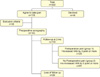
Between January 2009 and December 2010, 222 men had a vasectomy at our hospital, with 178 completing the preoperative questionnaire. Preoperative scrotal ultrasonography was performed in 144 patients who met the inclusion criteria. At 2 months after vasectomy, 114 (79.1%) of these men completed the follow-up questionnaire, scrotal ultrasonography, and semen analysis (Fig. 1). The average age of the 114 men was 36.3 years old (range, 29 to 53 years). At the scrotal ultrasonography at 2 months after vasectomy, epididymal thickening including tubular ectasia was noted in 110 men (96.5%). In all, 23 men (20.2%) described the new onset of scrotal discomfort after vasectomy. In the current study, new onset of scrotal discomfort after vasectomy was defined as having a postoperative visual analogue scale (VAS) score higher by 3 points or more than the preoperative VAS score. Subsequently, the subjects were analyzed separately and divided into two groups according to scrotal pain or discomfort.
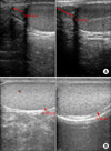
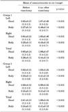
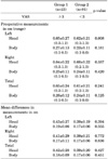
Epididymal measurements showed significant differences between before and after vasectomy (Table 1)(Fig. 2). As shown in Table 2, group 1 (n=23), who reported scrotal pain or discomfort, showed no significant differences in the maximal diameter of both epididymal heads when compared with group 2 (n=91), who had no scrotal pain or discomfort. Also, the width of both epididymal bodies did not differ significantly between the two groups. Spermatocele was detected in two cases in group 1. Hydrocele and varicocele were not detected. No significant differences in epididymal measurements were noted between the two urologists.
DISCUSSION
Vasectomy remains a common operation for urologists. Chronic testicular or scrotal pain is a recognized complication of vasectomy, but the exact incidence of such pain remains unknown [2]. Contemporary studies suggest that chronic postvasectomy pain syndrome (PVPS) is more common than originally thought. McMahon et al reported chronic testicular pain in 33% of men who had undergone vasectomy, which was troublesome in 15% and caused 5% to seek medical attention [2]. Choe and Kirkemo identified chronic scrotal pain in 18.7% of patients after vasectomy, and it had an adverse effect on their quality of life in 2.2% of those cases [8]. In our analysis, the incidence of chronic scrotal pain after vasectomy was 20.2%. The present results are similar to those previously reported by others. Scrotal pain after vasectomy has been referred to by many terms, including late postvasectomy syndrome, postvasectomy orchalgia, congestive epididymitis, and PVPS [1,9-11]. Currently, the syndrome is generally termed PVPS [12].
The cause of chronic PVPS remains controversial; one leading theory proposes that the obstruction and resulting dilatation of the epididymal duct produces interstitial fibrosis. It has also been suggested that pain results from perineural fibrosis and inflammation after the rupture of epididymal ducts, with extravasation of spermatozoa around the epididymal tubules and at the site of vasal transection. The nerves in these areas become densely encased in fibrous tissue, with distortion, angulation, and lymphatic infiltration [2]. It is unclear why some patients develop persistent symptoms, whereas others have only transient complaints. A different degree of local fibrosis after an inflammatory response and the varying extent of epididymal compression might account for the subjective experience of scrotal pain in some patients. West et al described the histological findings of epididymides in patients with scrotal pain after vasectomy as epididymal engorgement, complex cystic disease, and chronic epididymitis [13]. It is most likely that the ultrasonographic feature of tubular ectasia of the epididymis represents engorgement of the epididymal body from obstruction. Quantitative morphometric analysis of testicular histology in men after vasectomy showed dilatation of the seminiferous tubules, interstitial fibrosis, and reductions in the seminiferous cell population [14]. In appropriately selected patients, vasovasostomy can produce marked improvement or resolution of pain. This therapy has the obvious drawback of restoring fertility. Nangia et al reported that 69% of patients were pain-free after reversal, noting that the selection criteria for surgery are important to the outcome [15]. A careful preoperative evaluation should include serial physical examinations to confirm the site of persistent pain, consideration of a psychological evaluation to exclude somatization, and scrotal ultrasound to assess for occult pathology. Myers et al reported, in a small series, that 84% of patients with PVPS had complete resolution of pain after vasovasostomy [16]. Our study prospectively evaluated the scrotal pain and postvasectomy status on scrotal ultrasonography after vasectomy. It was hypothesized that chronic scrotal pain after vasectomy would be associated with obstruction of the epididymal duct. However, our results showed no significant differences in ultrasonographic features at 2 months after vasectomy between the two groups. Nevertheless, this is the first study that has described the relationship between ultrasonographic features and PVPS.
The differential diagnosis of scrotal pain after vasectomy must include nerve impingement or injury, varicocele, hydrocele, infection, testicular neoplasm, intermittent testicular torsion, inguinal hernia, referred pain, and psychogenic causes [4]. Although each of these is a potential cause of pain, most can be excluded by a thorough history, physical examination, and urine analysis [12]. The main limitation of the present study is the relatively short-term follow-up. For the present study, the follow-up duration has so far been restricted to 8 weeks. We are continuing to evaluate the patients at 6 months after vasectomy to obtain long-term results.
CONCLUSIONS
In summary, in our study with short-term follow-up, we found no statistically significant differences in the enlargement of the epididymis or scrotal ultrasonographic features between the scrotal pain and no pain groups. Therefore, causes of scrotal pain after vasectomy other than obstruction may need to be considered.
 XML Download