

 PDF
PDF ePub
ePub Citation
 Print
Print


Inguinal hernia is a common disorder that requires surgical management. Many organs can be associated with inguinal hernias, but bladder involvement is seen in less than 4% of cases [1,2]. The incidence may reach 10% among obese men who are 50 to 70 years old. Most cases are asymptomatic and are usually found incidentally during radiographic evaluation or at the time of herniorrhaphy [3]. Massive inguinoscrotal bladder hernia, also known as scrotal cystocele, is very rare [4]. In Korea, there was only one reported case of inguinoscrotal bladder hernia by Kim et al [5]. Here we report a case of massive inguinoscrotal bladder hernia in a 64-year-old man who presented with a scrotal mass. We also provide a review of the relevant literature.
CASE REPORT
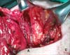
A 64-year-old man presented to our institution with an elevated serum prostate-specific antigen (PSA) level and a right scrotal mass. The PSA elevation had been found incidentally during a medical examination, and the right scrotal mass was first noticed 25 years previously. He had undergone right inguinal herniorrhaphy 30 years previously, and his additional comorbidities included hypertension. Physical examination revealed a large cystic mass in the right hemiscrotum. The patient reported no urinary symptoms or previous episodes of urinary retention. The scrotal mass was not reducible, but the mass was sometimes enlarged after sexual intercourse. The body mass index of the patient was 26.4 kg/m2. His PSA was 4.3 ng/ml, and his prostate volume was 28.8 cc on transrectal ultrasonography. On uroflowmetry, maximum flow rate (Qmax) was 40 ml/s and the voided volume was 280 cc. The uroflow curve was bell-shaped. A bladder scan revealed 8 cc of residual urine. The results of laboratory tests, including urinalysis and urine cultures, were normal. Ultrasonography of the testes revealed a large cystic mass in the right scrotum that was greater than 5 cm, without any solid portion or vascularity, and the connection to the pelvic cavity was not definite. Both testes and the epididymis were normal (Fig. 1). No further evaluation was performed, and the patient underwent surgical exploration. Under general anesthesia, a prostate biopsy was performed before surgical exploration. Bladder hernia was not considered preoperatively, and the right scrotum was explored. The large cystic mass was surrounded by fat tissue and was found to be connected to the pelvic cavity. Filling of the bladder revealed inguinoscrotal herniation of the bladder, and right inguinal exploration was performed. The bladder was dissected from the inguinal canal and was found to have directly herniated through the right margin of the rectus muscle (Fig. 2). Intraoperative findings demonstrated no evidence of bladder necrosis or hernia neck. The bladder was returned to its normal pelvic position without resection, and the inguinal floor and rectus margin were repaired by using mesh.
The patient had an uneventful postoperative course. Cystography performed 2 weeks after the operation demonstrated a normal bladder. Pathologic examination of the prostate revealed benign prostatic hyperplasia (BPH).
DISCUSSION
The bladder is involved in less than 4% of all inguinal hernias, and most cases are not diagnosed before surgical repair [1,6]. Most bladder hernias are direct, with a 70% male predominance, and most cases occur on the right side [7].
Although it is important to make the diagnosis preoperatively to reduce complications, less than 7% of bladder hernias are diagnosed before surgery: 16% are diagnosed postoperatively owing to complications, and the remaining cases are diagnosed perioperatively [2]. In the present case, the bladder hernia was diagnosed perioperatively, and the patient had no specific symptoms that prompted consideration of bladder hernia. Bladder hernias are usually asymptomatic but are often associated with intermittent swelling in the groin and significant lower urinary tract symptoms (LUTS) [3]. In cases of large inguinoscrotal bladder hernias, the patients typically present with two-stage micturition, involving spontaneous bladder emptying with a second stage of manual compression of the hernia [7,8]. The following are possible pathophysiologies of bladder hernias: bladder outlet obstruction, chronically distended bladder, decreased bladder tone, obesity, and weakness of the pelvic wall [4,6]. In this case, the patient had a history of herniorrhaphy 30 years before presentation, and a right scrotal mass was detected 5 years after the herniorrhaphy. In a previous small series of bladder hernias, one of four patients also had a history of herniorrhaphy several years before presentation [3]. In this case, the herniated bladder protruded through the right side of the rectus muscle. However, whether a history of herniorrhaphy affects the occurrence of bladder hernia is uncertain.
Inguinoscrotal bladder hernia can be subdivided into the paraperitoneal, intraperitoneal, and extraperitoneal type according to the relation with the parietal peritoneum [6,9]. The paraperitoneal type, in which the extraperitoneal portion of the bladder lies medially to the hernia sac, is the most common [6,9]. In our case, the bladder was herniated directly without being covered by the peritoneum, which can be classified as the extraperitoneal type.
The patient in the present case did not complain of LUTS, and the prostate was not sufficiently enlarged to cause bladder outlet obstruction. However, Kraft et al reported four cases of inguinoscrotal bladder hernia with significant LUTS [3]. Whether LUTS are caused by entrapment of the herniated bladder or bladder outlet obstruction is uncertain, but the authors suggested that a large component of the LUTS was related to inguinoscrotal bladder hernia because the LUTS resolved in all four patients within a few months of hernia repair. Along with BPH, bilateral hydronephrosis, with or without acute renal failure; lithiasis in the herniated bladder; vesicoureteral reflux; necrosis of bladder; and scrotal abscess can also be associated with inguinoscrotal bladder hernia [2]. In their review of the literature, Oruç et al found 13 cases (11.1%) with malignancy among 116 patients with bladder hernia [2]. Nine of those were bladder carcinoma, three were prostate carcinoma, and one was reported as a neoplasm. These findings suggest that evaluation for malignancy should not be delayed in suspicious cases because of the high ratio of malignant cases in patients with bladder hernia.
Radiologic diagnosis can be established by cystography, intravenous pyelography (IVP), computed tomography (CT) scan, or ultrasonography. A dumbbell-shaped bladder can be diagnostic on cystography, and the diagnostic triad for bladder hernia on IVP consists of lateral displacement of the distal third of one or both ureters, a small asymmetrical bladder, and incomplete visualization of the bladder base [6,10]. CT scan and ultrasonography, which are current investigative modalities for scrotal cystocele, can identify the anatomical deformity and any potential complications, such as bladder or bowel infarction [10]. In this case, the diagnosis of bladder hernia was missed preoperatively even though ultrasonography was used. However, bladder hernia should be in the differential diagnosis if fluid collection is identified in the groin on ultrasonography. Other diagnostic clues include tissue in the hernia canal and scrotum with similar echogenicity to bladder tissue, alteration in the dimensions of the inguinal contents before and after voiding, and a visible bladder connection [10].
The standard treatment of inguinoscrotal bladder hernia is either reduction or resection of the herniated bladder followed by surgical repair. Bladder resection is recommended only for cases with bladder necrosis, a hernia neck of less than 0.5 cm in diameter, a bladder diverticulum, or tumor in the herniated bladder [9]. When bladder resection is performed, the vesicoureteral junction should be identified to minimize ureteral injury. Bladder augmentation may be required at a later stage secondary to decreased bladder capacity [2]. Occasionally, patients may opt for conservative therapy such as watchful waiting or intermittent urethral catheterization [8].
Inguinoscrotal bladder hernia remains a rare condition, especially cases of massive scrotal cystocele. Often misdiagnosed, unrecognized bladder hernia can lead to injury of the bladder during surgery. Older age, male sex, and obesity are risk factors, and further evaluation should be considered in these high-risk patients if any of the suspicious symptoms or urological pathologies mentioned above are present.

 XML Download