

 PDF
PDF ePub
ePub Citation
 Print
Print


INTRODUCTION
Since its introduction by Clayman et al, laparoscopic surgery has enabled great progress in the area of urology, and less invasive techniques such as laparoendoscopic single-site surgery (LESS) and natural orifice transluminal endoscopic surgery (NOTES) have recently been introduced to clinical practice [1]. Minimally invasive procedures such as LESS and NOTES have an aesthetic advantage over conventional laparoscopic surgeries because they require fewer portals of entry, which leaves minimal to almost no evident surgical scars.
Laparoscopic partial nephrectomy is performed as the surgery of choice for small renal tumors with established evidence [2], and the surgical effectiveness and overall oncological outcomes of this procedure have been proved to be equal to those of conventional open surgeries for selected patients, even in the case of locally advanced renal tumors [3]. For radical nephrectomy, the LESS technique is currently being practiced in many institutions, and its effectiveness has already been established. However, in the case of partial nephrectomy, the LESS technique is not yet actively practiced in clinical fields owing to its technical difficulties.
We performed LESS partial nephrectomy in a porcine model with the objectives of overcoming the technical challenges of LESS and exploring the feasibility of the procedure from a technical viewpoint.
MATERIALS AND METHODS
Under the approval of the Seoul National University Institutional Animal Care and Use Committee (SNUIACUC), six partial nephrectomies were performed on bilateral kidneys of a 5-month-old swine that weighed 30 kg. Three operations were done on each kidney by a surgical staff consisting of two urologists with much experience in laparoscopic surgery (E1, E2) and two less-experienced urologists (B1, B2). A 5 mm flexible laparoscope was used for vision, and the choice of articulating needle driver, rigid needle driver, grasper, and dissectors was made according to the preference of each surgeon.
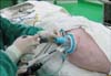
While under general anesthesia, the swine was placed in a lateral flank position. After a 2.5 cm skin incision was made at the umbilicus, the subcutaneous fat tissues were dissected and the peritoneum was opened. The Octoport™ wound retractor (type B, designed for a small wound) was put into the peritoneal cavity covering from the skin to the peritoneum. The Octoport™ wound retractor has 3 access ports, one 12 mm port and two 5 mm ports, and also has an additional gas inlet and gas outlet (Fig. 1). After applying the wound retractor, a 15 mmHg pneumoperitoneum was made with insufflation of CO2 gas. The renal hilum and Gerota's fascia were dissected to expose the renal capsule. After scoring around the imaginary tumor by use of monopolar electrocautery, a bulldog clamp was applied on the renal artery and parenchymal resection was done. Bilateral renal hilar dissections were done by E1. E1 performed a partial nephrectomy on the anterior side of the right kidney upper pole, E2 on the anterior side of the right kidney lower pole, and finally B1 on the posterior side of the right kidney lower pole, followed by B1's right nephrectomy. The swine was then repositioned to the opposite decubitus position. After repositioning, E1 performed a partial nephrectomy on the anterior side of the left kidney upper pole, E2 on the anterior side of the left kidney lower pole hilar area, and last, B2 resected the left kidney after performing partial nephrectomy on the posterior side of the left kidney lower pole. All operators used 3-0 Vicryl running sutures to repair the open calyx and for hemostasis of the resection bed and 1-0 Vicryl interrupted sutures for renorrhaphy, which was done with sliding knots only, without the use of any suture slip equipment such as Hem-o-lok (Teleflex Medical, Research Triangle Park, NC, USA) or Lapra-ty (Ethicon, Somerville, NJ, USA). During the calyx and parenchyma suture, articulating needle drivers were used alone for the repair of the resection margin of E1's first procedure and B2's procedure, and an articulating needle driver and rigid needle driver were used concurrently in all other cases.
RESULTS
All six partial nephrectomies were successfully performed without the need to introduce any additional ports, and all hemostatic procedures were successful in the repair of the vascular and collecting systems on the resection bed and renorrhaphy. Stable vital signs were maintained throughout the procedures, and there were no noticeable complications.
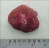
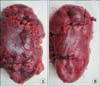
During the partial resection of the renal parenchyme, an articulating grasper and articulating cold scissors were used in all cases. The mean time for partial resection was 3.1 minutes (range, 2.5-3.3 minutes), and the mean resection times for the upper and lower pole areas were 3.4 (range, 3-4) and 2.8 (range, 2.5-3.3) minutes, respectively. The experienced (E1, E2) and less-experienced (B1, B2) surgeons' mean times for partial parenchymal resection were 2.9 minutes (range, 2.5-3.3 minutes) and 3.25 minutes (range, 2.5-4 minutes), respectively. The mean sizes of the resected parenchyma were 2.1 cm (range, 1.9-2.4 cm) and 1.5 cm (range, 1.4-1.6 cm) for the long diameter and depth, respectively.
Regarding warm ischemic time, E1 recorded 60 minutes on the anterior side of the right kidney upper pole and 35 minutes on the anterior side of the left kidney upper pole. E2 scored 30 minutes on the anterior side of the right kidney lower pole and 35 minutes on the anterior side of the left kidney lower pole around the hilar area. For B1 and B2, the warm ischemic times were 47 and 50 minutes for the posterior side of the right kidney lower pole and the posterior side of the left kidney lower pole, respectively. The shortest ischemic time, 30 minutes, was achieved by E2 by use of the early unclamping technique during renorrhaphy. The mean warm ischemic time was 42 minutes (range, 30-60 minutes); according to imaginary tumor location, it was 47.3 minutes (range, 35-60 minutes) for upper pole tumors and 36.7 minutes (range, 30-50 minutes) for lower pole tumors, respectively. The experienced (E1, E2) and less-experienced (B1, B2) surgeons' mean ischemic times were 38.8 minutes (range, 30-60 minutes) and 48.5 minutes (range, 47-50 minutes), respectively. The average number of sutures for renorrhaphy was 3.2 (range, 2-5).
DISCUSSION
The rising demand for minimally invasive surgical procedures and the development of surgical techniques to meet such a need has enabled urologists to apply the single-port surgery techniques to most laparoscopic surgeries in urology. The Cleveland Clinic has reported 100 surgical cases of LESS ranging from nephrectomy to even radical cystectomy [4]. However, there are relatively fewer successful reports on LESS in partial nephrectomy. Boylu et al reported 10 cases of partial nephrectomies using a porcine model [5]. At this point in clinical practice, there are only initial case reports of LESS in highly selected cases. Aron et al reported 4 human cases in highly selected patients, but they used an additional 2 mm grasper via a 2 mm Veres needle for the purpose of tissue traction [6]. Our study focused on evaluating in a porcine model the technical feasibility of using a pure LESS technique for performing partial nephrectomy.
In this study, all six partial nephrectomies were successfully done without any significant complications. However, there are some obstacles to performing LESS partial nephrectomy in humans. The most important one is the learning curve. One obvious difference between the LESS surgical technique and pure laparoscopy is the limitation in the movement of the instruments. That limitation makes the LESS surgery difficult and the learning curve for LESS surgery steep.
Three major procedures make it difficult to manipulate the surgical instruments properly in LESS partial nephrectomy. These are tumor resection, calyceal suture, and parenchymal suture. For tumor resection, it took only 2.9 minutes for the experienced laparoscopic surgeons, and even for the inexperienced surgeons, it took only 3.3 minutes with the use of the flexible instruments. However, sewing the open calyx and resected renal parenchyma was the most difficult procedure during LESS partial nephrectomy. To identify whether the suture technique used in pure laparoscopic surgery could also be adapted for LESS partial nephrectomy, the surgeons in this study did not use any laparoscopic clips, which can make suturing easier and faster. The most difficult procedure in LESS partial nephrectomy is parenchymal suture. Therefore, reducing the time for this procedure can shorten the warm ischemic time. Many researchers have tried various ways to reduce the warm ischemic time. We tried only interrupted parenchymal sutures with the sliding knot technique, which is simple and accurate but the most difficult and time-consuming technique, with the object of identifying the feasibility of this suturing technique for LESS partial nephrectomy. Therefore, it took from 27 minutes to 57 minutes for the calyceal and parenchymal sutures.
The articulating instrument is very useful during all of the procedures in LESS surgery, especially for triangulation and procuring enough space for the instruments to move freely in the restricted space. In our experience, it was convenient for the right-handers to use the articulating instrument for the right-side instrument, because the right-hand instrument usually performs the counter-traction to make the dissection easier in contrast with pure laparoscopic surgery. However, which hand should use the articulating instrument depends on the situation. Regarding the needle drivers, the articulating needle drivers with their flexible characteristics did not provide sufficient strength during sutures, which made the sewing procedures more difficult. Therefore, replacing the flexible needle drivers with rigid, conventional needle drivers became more convenient. When replacing the articulating needle driver with a rigid needle driver, the surgeons changed the right-hand instrument to the rigid needle driver, because all the surgeons in this study were right-handed. Also, we used right-angled forceps for the left-hand instrument and instead of using triangulation of the instruments we coordinated both instruments in a parallel position. During knot-tying, however, the instruments should be coordinated in a forward and backward direction, because there is no space for the instruments to move in a lateral direction as in pure laparoscopic surgery.
During the renal parenchymal suturing, we used early unclamping and the sliding knot technique to shorten the ischemic time. Describing our early unclamping technique in detail, first, an interrupted renal parenchymal stitch was made. After making a loose square knot, we converted the square knot to a sliding knot. Then two or three more stitches were made according to the size of the parenchymal resection. We made just one stitch fasten, unclamped the bulldog clamp, and finally fastened the other stitches. This early unclamping technique resulted in the shortest warm ischemia time of 30 minutes.
The limited warm ischemic time also makes LESS partial nephrectomy difficult. There are many controversies about warm ischemic time during partial nephrectomy. Shikanov et al reported in a multicenter study that ischemic time (mean ischemic time was 29 minutes) does not affect global renal function in the case of two normal functioning kidneys [7]. However, Becker et al reported in a mass analysis that in order to minimize parenchymal ischemic injury, the tumor should be removed within 20 minutes of warm ischemia, regardless of the surgical approach [8]. Large-scale institutions today score mean ischemic time around 30 minutes in the case of laparoscopic partial nephrectomy and it is assumed that warm ischemic time under 30 minutes is required in order to successfully apply LESS to partial nephrectomy in clinical practice [9-11]. In our study, the mean warm ischemic time was 42 minutes. The experienced urologists group scored an average of 38.8 minutes, and E2 achieved 30 minutes by using the early unclamping technique. However, considering that this study was performed with a porcine model, a longer warm ischemic time can be assumed in the case of a human kidney. Compared with the human body, the porcine model we used was smaller and had a smaller resected volume, which results in a shorter distance and smaller defect to repair. Furthermore, it is believed that it is easier to obtain hemostasis in a porcine model than in a human kidney because of the different cell biology, blood supply, and tissue textures. All the above factors will make the procedure of LESS partial nephrectomy more difficult in humans than in the porcine model. Nguyen and Gill reported halving the ischemic time from 31.1 to 13.9 minutes by using the early unclamping technique, although the technique was associated with a risk of bleeding and reclamping [12]. However, we can also infer that the early unclamping technique and dry and wet laboratory disciplinary efforts to handle the instruments more freely can reduce the warm ischemic time. Although Hem-o-lok (Teleflex Medical, Research Triangle park, NC, USA) or surgical clips such as Lapra-Ty (Ethicon, Cincinnati, OH, USA) are generally used in laparoscopic partial nephrectomy, our study used only sliding knots for suturing. Proper use of surgical clips may enable further decreases in ischemic time.
Another important fact that we found in this study was that there was no need for an additional trocar for LESS partial nephrectomy. All four surgeons could finish the LESS partial nephrectomy without any added trocars, even for right partial nephrectomies.
Boylu et al reported 10 consecutive LESS partial nephrectomies in porcine models performed by a single surgeon [5]. That study showed an overcoming of the learning curve with decreasing warm ischemic time. In our study, because of the limited number of cases and the many surgeons, the learning curve could not be assessed. But we can postulate that consecutive dry and wet laboratory discipline and experience would improve surgical technique, shorten the warm ischemic time, and result in the surgeon overcoming the learning curve.
CONCLUSIONS
LESS partial nephrectomy was successfully performed in a porcine model without significant complications. However, our study suggests that LESS for partial nephrectomy results in longer ischemic times than for conventional laparoscopic surgeries. Therefore, many dry and wet laboratory disciplinary efforts are needed to decrease the warm ischemic time and improve this surgical technique.
 XML Download