

 PDF
PDF ePub
ePub Citation
 Print
Print


Polyarteritis nodosa (PAN) is a rare, systemic necrotizing vasculitis that is generally restricted to small and medium-sized vessels. The incidence is about 0.4 to 1.0 per 100,000 and the disease is most prevalent in men aged 40 to 60 years [1]. In most cases, the etiology of PAN remains unknown, although it can be associated with hepatitis B and other viral infections. PAN can affect any organ system, but more commonly involves peripheral nerves, skin, and the gastrointestinal tract. Symptomatic testicular involvement can occur in around 18% of patients, and this frequency is highlighted by the fact that testicular involvement is one of the American College of Rheumatology (ACR) diagnostic criteria for PAN. Cranial nerve involvement is rare. Here we describe a patient in which bilateral testicular involvement was a presenting symptom and the diagnosis was made on the basis of testicular histopathology after the patient developed a seventh cranial nerve palsy.
CASE REPORT
A previously healthy 53-year-old Asian man presented to the emergency department with a 1-day history of severe bilateral testicular pain and swelling. He reported feeling generally unwell for 3 weeks with fatigue, myalgias, and intermittent fevers. He had no history of dysuria, hematuria, urethral discharge, prostatism, scrotal trauma, or surgery. On examination, he was febrile (37.9℃), his right eye was noted to be inflamed, and there was mild swelling of both ankles. There was bilateral enlargement and tenderness of the testes. Investigations revealed a hemoglobin level of 144 g/l (normal range: 135-180), white cell count of 14.5×10⁹/l (normal range: 4-11), neutrophils of 12.9×10⁹/l (normal range: 2-8), and C-reactive protein of 94 mg/l (normal range: <5). His bilirubin was 43 µmol/l (normal range: 3-18), GGT was 149 µ/l (normal range: 5-65), and albumin was 23 g/l (normal range: 35-46). Creatinine, ANCA, ANA, EBV serology, urinalysis, and testicular tumor markers were normal.
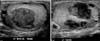
A computed tomography scan of the abdomen and pelvis was normal. Scrotal ultrasound revealed two avascular, intratesticular lesions of 2.3 cm and 2.1 cm in diameter on the left and a similar 2 cm lesion on the right (Fig. 1). A diagnosis of severe bilateral orchitis was made and the patient was treated with intravenous ampicillin and gentamicin but failed to improve. On the fourth day of the hospital admission, the patient developed left facial weakness and the CRP level increased further to 190 mg/l.
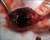
A diagnosis of vasculitis was suspected and the patient underwent a right testicular biopsy. Intraoperatively, more than one third of the testicle was necrotic with coagulated blood (Fig. 2). Histopathology demonstrated features of transmural inflammation and fibrinoid necrosis of medium-sized vessel walls, consistent with PAN (Fig. 3). Only one testis was explored and minimal debridement was done to conserve as much testicular tissue as possible. Postoperatively, treatment consisted of intravenous methylprednisolone and cyclophosphamide 500 mg weekly for 6 weeks followed by oral prednisone and cyclophosphamide maintenance. The testicular pain was resolved after 5 days and the left seventh nerve palsy was resolved after 3 months. Five months after disease onset he was asymptomatic and his CRP was 0.5 mg/l with prednisone 5 mg/day and cyclophosphamide 500 mg/wk. After 6 months of follow-up, no relapse had occurred and his testosterone level was in the low normal range.
DISCUSSION
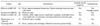
PAN is a multisystem necrotizing vasculitis that is characterized by segmental transmural inflammation of small or medium-sized arteries with resultant tissue necrosis. PAN may present as either a limited or systemic disease and may be either primary or secondary when associated with hepatitis B virus infection, a connective tissue disease, or leukemia. Testicular involvement is one of the ACR diagnostic criteria for PAN (Table 1) [2]. Although isolated vasculitis of the testis is uncommon, involvement of the testis is reported in up to 86% of systemic PAN in autopsy studies [3]. Uncommonly, testicular involvement is the initial manifestation of systemic disease and the clinical presentation may include pain, swelling, atrophy, or a palpable mass. PAN is often diagnosed late and may be mistaken for acute infection, torsion, or tumor. Systemic manifestations such as fever, fatigue, and weight loss further confound the diagnosis. Unfortunately, imaging cannot reliably differentiate vasculitis from a neoplasm in the testis. Both conditions may present as a mass lesion and reveal multiple hypoechoic areas on scrotal ultrasound. Definitive diagnosis of PAN requires tissue biopsy.
Our case was interesting because the patient also developed a unilateral facial nerve palsy. Neurologic abnormalities are reported to occur in 25% to 50% of patients with PAN [4]. Mononeuritis multiplex is the most common manifestation, with cranial nerve palsies present in less than 2% of cases. The oculomotor (III), trochlear (IV), abducens (VI), facial (VII), and acoustic (VIII) nerves are affected most often.
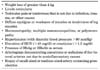
With the introduction of corticosteroids and immunosuppressants such as cyclophosphamide, the 5-year survival rate has increased from 13% to 80% [5]. With evidence of neurological involvement, our patient was started on high-dose intravenous methylprednisone and cyclophosphamide with good effect. In this case, we also report successful conservative management with normal endocrine testicular function (Table 2) [6-9]. The facial weakness resolved after 3 months.
To our knowledge, this is the first reported case of PAN presenting with bilateral testicular swelling with the subsequent development of a facial nerve palsy. This case highlights that the diagnosis of PAN needs a high index of suspicion in presentations of testicular swelling accompanied by systemic symptoms. An expedited diagnosis will permit early instigation of treatment and could prevent the extent and distribution of other end-organ damage.

 XML Download