

 PDF
PDF ePub
ePub Citation
 Print
Print


INTRODUCTION
Various causes determine the formation of ureter calculi, and genetic factors are known to play a role. Studies have tried to identify genes related to ureter calculi in an effort to clarify the cause of urolithiasis and to advance the diagnosis and treatment of urolithiasis [1,2]. In other studies, the use of single-nucleotide polymorphisms (SNPs) associated with genetic diseases has been fruitful in identifying candidate disease genes. Recent genetic advances in urolithiasis indicate the potential of a new approach towards the gene polymorphism [3-7].
Interleukin-1β (IL-1β) is a potent proinflammatory agent that can induce bone resorption, osteoclast formation, and hypercalciuria. The calcium-sensing receptor (CaSR) regulates cellular calcium homeostasis and controls parathyroid hormone secretion that is controlled by urinary calcium absorption. Urokinase functions to degrade the organic matrix of nascent urinary stones, preventing their full formation and growth. Hypothesizing that these genes might be crucial in urolithiasis, the present study examined gene polymorphisms in IL-1β, CaSR, and urokinase in Korean urolithiasis patients and healthy controls.
MATERIALS AND METHODS
1. Subjects
From January 2007 to December 2008, patients who had a confirmed diagnosis after a radiological exam, such as computed tomography, intravenous pyelography, or kidney ultrasonography, and who were treated for urolithiasis with open surgery, ureterorenoscopic lithotripsy, extracorporeal shock wave lithotripsy, or conservative treatment at Chung-Ang University Hospital were involved in this study. Informed consent for genetic testing was given by those who agreed to participate. The control group consisted of patients displaying normal urinalysis findings from the health screening, absence of stones as identified by ultrasonography, and no history of urolithiasis. Genetic testing was performed as described below to detect polymorphisms in IL-1β (484 urolithiasis patients, 208 controls), CaSR (433 urolithiasis patients, 197 controls), and urokinase (370 urolithiasis patients, 167 controls). The study protocol was approved by the Chung-Ang University Institutional Review Board.
2. Methods
1) Genomic DNA isolation
Genomic DNA was isolated from peripheral blood leukocytes in EDTA-containing tubes. All processes were done according to the manufacturer's instructions by use of the Wizard Genomic DNA Purification Kit (Promega, Madison, WI, USA).
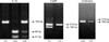
2) Amplification and confirmation of each mutation (Fig. 1)
(1) IL-1β T/C
The extracted DNA was amplified by polymerase chain reaction (PCR) with the forward primer 5'-G CCTGAACCCTGCATACCGT-3' and the reverse primer 5'-GCCAATAGCCCTCCCTGTCT-3'. PCR procedures were carried out in a total volume of 20 µl containing genomic DNA, each primer, and Taq DNA polymerase. PCR amplification was performed in a programmable thermal cycler GeneAmp PCR system 9700 (Applied Biosystems, Foster City, CA, USA). The cycling conditions for IL-1β T/C polymorphism were 1 cycle at 95℃ for 5 minutes; 35 cycles at 94℃ for 30 seconds, 56℃ for 30 seconds, and 72℃ for 30 seconds; and a final cycle at 72℃ for 10 minutes. Then, 5 µl of the product was loaded onto 3% agarose gel plates containing ethidium bromide for electrophoresis. The 155-bp PCR product was mixed with 10 U/µl of AvaI restriction enzyme (Enzynomics, Daejeon, Korea) and the restriction buffer according to the manufacturer's instructions. The thymine in the wild-type sequence is replaced by cytosine as the polymorphic mutation, generating a recognition site for AvaI. The resulting digestion generates 88-bp and 68-bp fragments.
(2) CaSR: A986S
Primers were designed to amplify the region containing the A986S polymorphism (forward primer 5'-CTTTGATGAGCCTCAGAAGAGC-3' and reverse primer 5'-ACAACTCTTCAGGGTCCTCC-3'). The PCR cycling condition for CaSR gene polymorphism was 1 cycle at 95℃ for 5 minutes; 35 cycles at 94℃ for 30 seconds, 58℃ for 30 seconds, and 72℃ for 45 seconds; and a final cycle at 72℃ for 7 minutes. The 218-bp PCR product was mixed with 20 U/µl of SacI restriction enzyme (Enzynomics) and the restriction buffer according to the manufacturer's instructions. SacI would recognize a recognition site in the mutant allele producing fragments of 218-bp and 198-bp.
(3) Urokinase 3' untranslated region (UTR)
The extracted DNA was PCR-amplified by using the forward primer 5'-C CGCAGTCACACCAAGGAAGAG-3' and the reverse primer 5'-GCCTGAGGGTAAAGCTATTGTCGTGCAC-3'. The PCR cycling condition for the urokinase 3' UTR polymorphism was 1 cycle at 94℃ for 5 minutes; 35 cycles at 94℃ for 30 seconds, 58℃ for 30 seconds, and 72℃ for 40 seconds; and a final cycle at 72℃ for 7 minutes. The 210-bp PCR product was mixed with 20 U/µl of ApaL1 restriction enzyme and the restriction buffer according to the manufacturer's instructions. Polymorphism would generate an enzyme recognition site, and digestion would produce fragments of 185-bp and 25-bp.
3) Stone metabolic study
IL-1β, CaSR, and urokinase polymorphisms were determined in the urolithiasis patients. Each genotype was classified into groups and the average sodium, calcium, citrate, and oxalate concentrations in urine collected for over 24 hours were determined, as was the 24-hour urine volume.
4) Statistics
The chi-square test was used for the statistical analysis of gene frequency and allele frequency, and one-way ANOVA was used for the statistical analysis of metabolic factors between the three genes. SPSS ver. 12.0 (SPSS Inc., Chicago, IL, USA) was used, with p-values less than 0.05 considered significant.
RESULTS
1. Genotype frequency
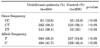
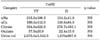
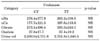
In urolithiasis patients, the C/C, C/T, and T/T IL-1β gene polymorphisms were detected in 61 (12.6%), 282 (58.3%), and 141 patients (29.1%), respectively. The respective proportions in the normal controls were 33 (15.9%), 123 (59.1%), and 52 patients (25%) (Table 1). In urolithiasis patients, the T/T and T/t CaSR gene polymorphisms were detected in 415 (95.9%) and 18 patients (4.1%), respectively. The respective proportions in the normal controls were 191 patients (97%) and 6 patients (3%) (Table 2). In urolithiasis patients, the C/T and T/T urokinase gene polymorphisms were detected in 212 (57.3%) and 158 patients (42.7%), respectively. The respective proportions in the normal controls were 104 (62.3%) and 63 patients (37.7%) (Table 3). No statistically significant differences were evident between the patient groups and the control groups for any of the genes.
2. Allele frequency
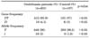
The C and T IL-1β allele frequencies in the 484 urolithiasis patients were 404 patients (41.7%) and 564 patients (58.3%), respectively. The respective proportions of the 208 control patients were 189 patients (45.4%) and 227 patients (54.6%). The T and t CaSR allele frequencies in 433 urolithiasis patients were 848 patients (98%) and 18 patients (2%). The respective proportions of the 197 normal controls were 388 patients (98.5%) and 6 patients (1.5%). The T and C urokinase allele frequencies in the 370 urolithiasis patients were 528 patients (71.3%) and 212 patients (28.7%), respectively. The respective proportions in the 167 normal controls were 230 patients (68.9%) and 104 patients (31.1%). No statistically significant differences were evident between the patient groups and the control groups for any of the genes.
DISCUSSION
Genetic factors influence the formation of urinary calculi. For example, the formation of urinary calcium stones, urinary calcium and hydroxyl concentrations, crystallization growth factors, and inhibitory factors are affected by genetic factors [8]. In addition, calcium oxalate stones have been linked to a polygenic defect [2]. Also, there is a report that hypercalciuria and urolithiasis are related. However, genetic details of hypercalciuria and urolithiasis patients are unclear; thus, it remains unresolved whether one or several genes are involved in calculi formation [8].
For a disease such as urolithiasis, which results from the complex interaction of multiple factors, determination of SNPs is useful to identify disease-related gene(s). The complete roster of polymorphic markers must be resolved with the need to identify 90% or more of the relevant SNPs. SNPs differ appreciably with race, although about 30% are common between all races. Much SNP research has been done in other countries, but none in Korea. In the present study, we aimed to determine SNPs in Korean patients.
IL-1β functions to localize the bone marrow macrophage to the site of inflammation, to stimulate differentiation of macrophages into osteoclasts by activation around the bone, and to cause hypercalciuria [9]. Similar to the present results, a study of IL-1β polymorphism conducted in China on patients with calcium oxalate stones did not find evidence of a statistically significant association of the polymorphism with the patient group [10]. A study in India reported a significantly higher risk for urolithiasis in patients with a combination of high IL-1β and low IL-1 receptor antagonist genotypes [11]. This is consistent with the aforementioned racial difference in gene polymorphism. It seems that research on genetic factors to determine why there are low relations between IL-1β and urolithiasis in Asians is needed. In the present study, we compared the laboratory results of 24-hour collected urinary sodium, calcium, hydroxyl, and citric acid and urine volume according to each genotype. No significant differences were revealed. CaSR gene inactivation caused by mutations is associated with diseases characterized by hypocalciuria and hypercalcemia because gene inactivation causes changes in parathyroid hormone secretion and calcium reabsorption of tubular cells. CaSR gene mutation also seems to be the cause of idiopathic hypercalciuria, which is a risk factor for calcium renal stone formation and is characterized by an increase in calcium excretion even when normal plasma calcium ion is maintained [10,12,13]. A study from Brazil reported that CaSR polymorphism was overrepresented in hypercalciuric stone formers but that urinary calcium excretion was not associated with polymorphism [14]. Presently, CaSR gene polymorphism analysis did not reveal genotype differences between urolithiasis patients and normal controls. When allele frequency was classified into T and t, no statistically significant difference was found, in agreement with the study of Italian urolithiasis patients (although statistical differences in CaSR mutations and hypercalciuira were reported) [15]. Likewise, the laboratory results of 24-hour urinary sodium, calcium, hydroxyl, and citric acid and urine volume were compared according to each genotype, and again no significant differences were revealed. Urokinase was originally isolated from human urine, but is present in several physiological locations, such as the bloodstream and the extracellular matrix. The primary physiological substrate of the enzyme is plasminogen, which is an inactive zymogen form of the serine protease plasmin. It is assumed that urokinase breaks the organic matrix of nascent urinary stones, preventing their complete formation and growth. In a study of urolithiasis urokinase gene polymorphism conducted on patients in China, T allele carriers had a statistically higher incidence of calcium oxalate stones [16]. A study from India also reported a significant difference in the distribution of the urokinase gene C/T polymorphism between patients with stones and controls [17]. In the present study, the urokinase gene polymorphism analysis showed no genotype differences between urolithiasis patients and normal controls in the prevalence of the C and T alleles, which may reflect racial differences. Likewise, there were no statistically significant differences in urine parameters.
 XML Download