

 PDF
PDF ePub
ePub Citation
 Print
Print


INTRODUCTION
Many factors are involved in the development, advancement, and metastasis of malignant tumors. Recently, there has been increasing attention paid to the interaction between cancer cells and the adjacent normal cells. Cell binding among normal cells connects the cells to each other and induces a stable interrelation, and zonula adherens, desmosomes, zonula occludens, and the tight junction are involved [1]. The tight junction consists of three integral transmembrane proteins: occludins, claudins, and junctional adhesion molecules [1-3]. Claudin, one of the main proteins that form epithelial tight junctions, was first discovered in 1998, and 24 types have been reported to date [4]. The up-regulation or down-regulation of claudin is known to be related to the carcinogenesis and metastasis of various cancers, such as stomach cancer, colon cancer, and pancreatic cancer [5-7]. However, studies of the clinical correlation between various cancers and the expression of claudin have shown different results. Thus, studies are actively being conducted to confirm a clear correlation.
In this study, we verified the immunohistochemical expression level of claudin-1 and claudin-7 with clear cell RCC and compared the clinicopathologic prognostic factors (sex, age, BMI, tumor size, TNM stage, Fuhrman nuclear grade, postoperative distant metastasis, and cancer-specific survival) to investigate the significance of claudin-1 and claudin-7 expression.
MATERIALS AND METHODS
1. Subjects
The subjects of this study were the 119 patients whose diagnosis of clear cell RCC was confirmed among the 154 patients who underwent radical nephrectomy or partial nephrectomy for RCC in our hospital from January 2000 to December 2007. After performing immunohistochemical staining of the clear cell RCC tissue for claudin-1 and claudin-7, we investigated whether the claudin was expressed individually and analyzed its correlation with sex, age, BMI, tumor size (<4 cm, 4-7 cm, >7 cm), TNM stage, Fuhrman nuclear grade, postoperative distant metastasis, and cancer-specific survival.
2. Immunohistochemical staining
A pathologist researched the H&E staining samples of the subjects, marked the representative tumor region, and took a core with a 3 mm diameter from the corresponding paraffin block to prepare the tissue microarray (TMA). The TMA paraffin block was sectioned into slices of 4 µm thickness and they were attached to a slide. Then, they were soaked in xylene for 5 minutes three times for paraffin removal and washed with distilled water after a moisturizing process. The citrate buffer solution (pH 6.0) was used for the pretreatment to recover the antigenicity. The staining was performed by using UltraVision LP detection (Thermo Scientific, Freemont, CA, USA) and Autostainer360 (Lab Vision, Fremont, CA, USA). The primary antibody was diluted with Claudin-1 (ab15098, Abcam, Cambridge, UK) and Claudin-7 (ab27487, Abcam, Cambridge, UK) at ratios of 1:200 and 1:500, respectively, and underwent reaction for 40 minutes at room temperature. Mayer's hematoxylin was used as the contrast staining, and an optical microscope was used for observation.
4. Statistical analysis
Fisher's exact test and linear by linear association were used to examine differences in the expression of claudin-1 and claudin-7 depending on the clinicopathological findings of the clear cell RCC. Kaplan-Meier analysis was used for the survival analysis. p-values of less than 0.05 were considered statistically significant. We used the statistical program SPSS ver. 17.0 (SPSS Inc., Chicago, IL, USA).
RESULTS
1. Clinicopathologic characteristics
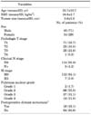
Clear cell RCC was found in a total of 119 subjects including 85 males (71%) and 34 females (29%). The patients' mean age was 55.7±10.7 years. The mean BMI was 24.6±2.7 kg/m2, and the mean tumor size was 5.6±2.8 cm.
The number of patients with preoperative distant metastasis was 7. The metastasis site was lung, liver, brain, and bone. No cases received neoadjuvant therapy, and target therapy was done in all cases after radical nephrectomy. The mean postoperative distant metastasis period was 21.7 months in the patients without preoperative distant metastasis, and 14 patients died from cancer-related events.
The clinicopathologic characteristics such as tumor stage, lymph node metastasis, Fuhrman nuclear grade, preoperative distant metastasis, and postoperative distant metastasis are shown in Table 1.
2. Correlation between expression of claudin-1 and clinicopathologic parameters
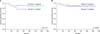
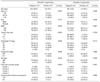
Claudin-1 was expressed in 18 cases (15.1%). The claudin-1 expression increased with age (p=0.007), but no correlation was found with sex or BMI (p=0.777 and p=0.559, respectively). The claudin-1 expression was significantly higher with larger tumor size (p=0.001), higher pathologic T stage (p=0.009), preoperative distant metastasis (p=0.035), and higher Fuhrman nuclear grade (p=0.004). Among the patients without preoperative distant metastasis, the claudin-1 expression was higher in the cases in which postoperative distant metastasis was present (log-rank test, p<0.001) (Table 2). However, there was no significant difference in the claudin-1 expression according to cancer-specific survival (p=0.110) (Fig. 3).
3. Correlation between expression of claudin-7 and clinicopathologic parameters
Claudin-7 was expressed in 31 cases (26.1%). The claudin-7 expression was significantly higher with higher Furhman nuclear grade (p=0.031). However, the claudin-7 expression was not correlated with age, sex, BMI, tumor size, TNM stage, or the existence of postoperative distant metastasis (all p>0.05) (Table 2). There was no significant difference between the claudin-7 expression and cancer-specific survival (p=0.662) (Fig. 3).
DISCUSSION
RCC is the most common primary malignant tumor in kidneys. The World Health Organization (WHO) classifies the malignant tumors in kidneys into 10 types, among which clear cell RCC, papillary RCC, and chromophobe RCC are the most common subtypes, accounting for about 85% to 90% of the total RCC cases [8,9].
Pathologic classification, TNM stage, and clinical opinion of RCC are known as important factors in the patient's prognosis [10]. Additionally, various factors in prognosis have been studied recently, such as the expression of claudin and its value as a prognostic factor. The loss of intercellular adhesion in tumor cells is known to be an important process in the intrusion and metastasis of tumor cells to the peripheral matrix [11]. Cell binding induces cells to interconnect with each other and have stable interrelations, and zonula adherens, desmosomes, zonula occludens, and tight junction are involved in this interaction [1]. Tight junctional proteins are located in the apical and basolateral sections of cells, which play an important part in the maintenance of polarity of cells, control of paracellular transportation, and barrier functions. As a result, cell binding represses the diffusion of the water and ions surrounding the cells as well as the internal macromolecules [12], controls the osmotic force, affects cell movement [1,13], and maintains homeostasis and cell polarity [2]. Moreover, cell binding is known to be involved in cell growth, differentiation, and control of other cellular functions.
Claudin, one of the integral transmembrane proteins that constitute tight junctions, is the main protein that forms the epithelial tight junction, and 24 types have been reported to date [4]. The expression of claudins is known to be related to the carcinogenesis of various cancers: the low expression of claudin-1 and claudin-5 was associated with a Gleason score of 7 or higher and a high prostate-specific antigen value in prostate cancer [14]. Claudin-1 was shown to be under- expressed in the tissues of prostate cancer, breast cancer, and thyroid cancer [15-17], and claudin-1 expression was lost in hepatocellular carcinoma [18].
Although claudin expression varies depending on the tumor, RCC has not been studied much, and only a few articles are available about its clinical prognosis in particular. Osunkoya et al and Hornsby et al reported that claudin-7 expression was enhanced in chromophobe RCC [19,20], and Fritzsche et al reported that claudin-1 expression was found in a small number of clear cell RCC patients but that cancer-specific survival was reduced in those patients [21]. In our study, claudin-1 expression was found in 15.1% of clear cell RCC cases, and it was higher with higher age, larger tumor size, higher T stage, preoperative distant metastasis, higher Fuhrman grade, and recent postoperative distant metastasis. By contrast, claudin-7 expression was found in 26.1% of clear cell RCC, and it was higher only with higher Fuhrman grade.
Because claudin exists at the tight junction between the cells and it maintains cellular polarity, serving as a wall, a reduction in claudin expression is anticipated as a tumor develops. However, according to various clinical results, claudin expression varies depending on the subtypes and the kinds of cancer [14-21]. Regarding the cause of such a contradiction among results, the protein level might have been increased as a response to the lost cellular cohesion during the malignant degeneration process or by the amplification due to mutation of the claudin gene [22]. Also, in our study, high stage and Fuhrman grade as well as distant metastasis were found in the cases in which the claudin expression was increased. The accurate role played by claudin in malignant cells should be investigated in future studies.
In the present study, we found that the correlation between the expression of claudin-1 and claudin-7 and cancer-specific survival was insignificant (p=0.110 and p=0.662, respectively). However, because there is a report that cancer-specific survival was reduced in patients in whom claudin-1 was expressed [21], and only a limited number of studies have been conducted on the relationship between claudin and cancer-specific survival until now, more studies should be carried out in the future to assess the correlation between claudin and cancer-specific survival.
A limitation of this study is that we had a small number of cases of other subtypes except clear cell RCC, such as papillary RCC and chromophobe RCC, and thus the expression and clinical characteristics of claudin-1 and claudin-7 were not investigated with respect to the other subtypes. Comparison of the claudin-1 and claudin-7 expression among different RCC subtypes needs to be carried out in the future with a larger number of cases.
CONCLUSIONS
In clear cell RCC, the claudin-1 expression was higher with higher age, larger tumor size, higher T stage, preoperative distant metastasis, and higher Fuhrman nuclear grade. When the claudin-1 expression was positive, the risk of postoperative distant metastasis was increased. The expression of claudin-7 was higher in relation to only a higher Fuhrman nuclear grade. Claudin-1 and claudin-7 were not correlated with cancer-specific survival. In clear cell RCC, the claudin-1 expression may be valuable as a clinically meaningful prognostic factor of postoperative distant metastasis.


 XML Download