

 PDF
PDF ePub
ePub Citation
 Print
Print


INTRODUCTION
Since percutaneous nephrolithotomy (PCNL) was first introduced in 1976, it has been recognized as a treatment for large-sized and lower pole renal stones or renal stones that cannot be dealt with by extracorporeal shock wave lithotripsy or ureteroscopic ureterolithotomy. PCNL has conventionally been conducted in the prone position [1]. However, prone position PCNL has several disadvantages [2]. Patients with cardiopulmonary problems, obesity, or skeletal deformity can show circulatory or ventilatory difficulties [3]. In addition, early intervention is difficult in cases of emergency following circulatory and ventilatory difficulties. Although it rarely occurs, vision loss can result from increased intraocular pressure [4]. Changing the position of a patient after anesthesia is an inconvenient and time-consuming procedure that can lead to more anesthesia-related morbidity. Because of these disadvantages, PCNL has been performed in the flank position, which is frequently used for renal surgery and is familiar to urologists at our institution. Flank PCNL can shorten the operation preparation time, and the shorter time reduces the risks associated with anesthesia. This study reports the peri-operative outcomes and learning curve for flank PCNL on the basis of a single surgeon's experiences.
MATERIALS AND METHODS
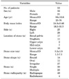
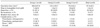
This study investigated 53 patients undergoing flank PCNL for renal stones at our institution from April 2008 to September 2010. Demographic and clinical characteristics of the patients are shown in Table 1. Stones in the renal pelvis, calyx, and staghorn were found in 17 (32.0%), 25 (47.2%), and 11 patients (20.8%), respectively. Mean stone size was 3.2 cm (range, 1.3-7.1 cm) and the largest renal stone was a 7.1 cm staghorn stone. Stones were irregular in most patients (27, 50.9%) and multiple stones were found in 23 cases. Radiopaque stones and radiolucent stones were found in 49 (92.5%) and 4 patients (7.5%), respectively.
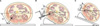
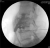
After general anesthesia, PCNL was performed in the flank position without an indwelling occlusion catheter in the lithotomy position. For renal access, the ipsilateral operating table was tilted by 20 degrees for lateralization of the lower calyx to make the puncture needle nearly vertical to the operating table (Fig. 1). The skin puncture for renal access was performed just below the lateral end of the 12th rib. Renal access was obtained through the appropriate renal calyx by a puncture needle by use of ultrasonography and fluoroscopy guidance. After dye injection, accurate targeting was checked by using fluoroscopy (Fig. 2). A guidewire was inserted with a ureteral balloon dilation catheter (UroMax Ultra™ High Pressure Balloon Catheter, Boston Scientific, Natick, MA, USA) during renal access and the tract was dilated. A 30 F Amplatz® working sheath (Boston Scientific) was inserted. After accurate renal access was checked, the ipsilateral operating table was lowered by 20 degrees to obtain the 30-degree flank position. Stones were then removed following the conventional PCNL technique.
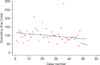
To examine therapeutic effect, stability, and the learning curve for flank position PCNL, mean operative time, stone-free rate, drop in hemoglobin level, length of hospital stay, complications such as blood transfusion and prolonged fever, and need for additional procedures after the surgery were compared among three groups obtained by dividing the 53 cases in chronological order. Stone-free rate was defined as the proportion of cases with residual stone(s) less than 5 mm on kidney, ureter, and bladder (KUB) X-rays on the first postoperative day. Noncontrast computed tomography (CT) was conducted to detect radiolucent stones and other complications such as hematoma and urinoma. ANOVA was used for comparison, and statistical analysis was done by using SPSS ver. 16.0 (SPSS Inc., Chicago, IL, USA). Data are expressed as mean±standard deviation with p<0.05 considered significant.
RESULTS
The mean operation time was 97.3±43.1 minutes. The mean operation time gradually decreased as the surgeon gained experience (Fig. 3). In particular, there was significant improvement in operation time in group 3. No statistically significant difference was found between groups 1 and 2 (p= 0.908), whereas group 3 showed a significantly decreased mean operation time compared with group 2 (group 2=112.1±52.7 min, group 3=72.2±24.1 min, p=0.019). The comparison of mean time between groups 1 and 2 combined and group 3 also revealed a statistically significant difference (group 1+2=109.1±45.2 min, group 3=72.2±24.1 min, p=0.003).
The mean operation time, drop in hemoglobin level, stone-free rate, re-treatment, length of hospital stay, and complication rate were compared for each cohort of patients as shown in Table 2. There was no significant difference in length of hospital stay among the groups (p=0.131). The overall stone-free rate was 64.2% for all procedures (range, 61.1-76.5% among the groups) (Table 2). There was no significant difference in the stone-free rate among the groups (p=0.88).
Each of the three groups had one case of re-treatment (ureteroscopic ureterolithotomy for one and extra shockwave lithotripsy for two patients). Drop in hemoglobin level and the frequency of blood transfusion slightly decreased as the surgeon gradually gained experience, but the difference was not statistically significant (group 1 vs. 2, p=0.520; group 2 vs. 3, p=0.343, respectively). There was no injury to the bowel or renal vessels, and no other major complications occurred following the procedure. Two patients experienced fever (defined as body temperature >38℃).
DISCUSSION
Since the first renal stone was removed via a nephrostomy tract by Fernström and Johansson in 1976 [1], PCNL has been accepted as a treatment for large-sized and lower pole renal stones and renal stones that are not easily addressed by extracorporeal shock wave lithotripsy or ureteroscopic ureterolithotomy. PCNL conventionally proceeds in the prone position [5].
Prone position PCNL provides a larger surface area for renal access and a wider space for instrument manipulation.
However, PCNL in the prone position has several disadvantages [6,7]. It can result in patient discomfort and lead to circulatory and ventilatory difficulties. According to the review by Edgcombe et al, complications related to anesthesia in the prone position can also occur [8]. First, the change in position from supine to prone can lead to carotid artery injury or occlusion of vertebral arteries due to excessive neck movement. Second, pressure caused by the chest roll can increase venous pressure, and if this occurs in the setting of mild arterial hypotension, it can provoke decreased spinal cord perfusion pressure or ischemia. In addition, contact dermatitis, tracheal compression, salivary gland swelling, mediastinal compression, and gas embolism can follow pressure injury to dependent parts of the body in the prone position. Although rare, Walick et al reported that vision loss can occur as the result of increased intraocular pressure during operation in the prone position [9]. In the prone position, the forehead is padded conventionally, but if the padding compresses the eyeballs, central retinal artery thrombosis can result in blindness.
Because PCNL is an excellent modality for removal of renal stones, supine position or lateral position PCNL has been tried to overcome the limitations of prone position PCNL. Falahatkar et al randomly divided 80 patients into two groups of 40 and conducted PCNL in the supine and prone positions, respectively [10]. After an indwelling ureteral catheter was placed in the lithotomy position, one group underwent PCNL in the supine position and the second group in the prone position. The mean operation time was significantly shorter in the supine position PCNL group (74.7±25.1 min) than in the prone position group (106.87±17.5 min) (p<0.0001). There was no significant difference in stone-free rates with 80% and 77.5% in the prone and supine position PCNL groups, respectively. Mean hospital stay and bleeding requiring transfusion were also not significantly different between the two groups. No colon injury was observed in either group. Rana et al conducted tubeless PCNL in 184 patients in the supine position with 20-degree rotation using a towel roll below the ipsilateral flank [11]. Mean procedure time was 65 min (range, 45-110 min) and total stone clearance was achieved in 154 patients. No vascular or splanchnic injury or bowel transgression was found. Karami et al performed PCNL with ultrasound-guided renal access in the lateral decubitus flank position in 40 patients with renal stones >2 cm [12]. All 40 patients had successful access and the complete stone clearance rate was reported at 85%. Their mean operative time was 45 min (range, 32-75 min), and there was no visceral injury or bleeding that required transfusion.
Concerning colon injury during renal access, the supine position or flank position has been found to be more advantageous than the prone position. LeRoy et al reported that the probability of colon injury in the prone position was high among patients with advanced age and horseshoe kidney [13]. Because elderly patients have little perinephric fat, the colon is located in an inadequate posterior position in many cases. In addition, patients with horseshoe kidney show a high probability of posterior colon displacement due to the defect in the normal lateroconal fascia resulting from the retroperitoneal abnormality. This increases the probability of colon injury in the prone position [13]. Rodriguez et al claimed that in the prone position, the colon was located in the lateral surface of the kidney and while in the supine position it fell anteromedially, which reduced the probability of colon puncture [14]. By use of computed axial tomography (CAT) imaging in a controlled prospective study, Hadar and Gadoth reported that the rate of retrorenal colon was higher in the prone position (4.7%) than in the supine position (1.9%) [15]. Taking this background into account, our institution has conducted flank PCNL since April 2008.
Compared with previously reported supine position PCNL, the flank PCNL performed at our institution is similar. In most cases of supine PCNL, the patient was rotated by about 20 to 30 degrees with a towel roll. This study also used the lateral position with the operating table tilted only for renal access and then a supine position with a slope of 30 degrees (Fig. 1). Jeong et al reported the initial experience of flank PCNL conducted at our institution. Renal stones were removed effectively and safely and the learning curve showed surgical competence with accumulation of experience by the surgeon [16].
Two main questions can be raised by surgeons who are newly learning flank PCNL. The first is whether renal access can be conducted safely and accurately in the flank position. Flank PCNL has a wider surface area for the choice of puncture site compared with the supine or true lateral decubitus position. There is also less difficulty of access caused by the rib cage compared with supine or prone position PCNL. The normal kidney forms an angle of 50 degrees with the coronal axis of the body. In the flank position, renal access is conducted by rotating the ipsilateral operating table by around 20 degrees to lateralize the lower calyx so that the puncture needle is nearly vertical to the operating table. Puncture at 90 degrees results in less difficulty of access caused by the rib compared with the prone position. A mobile kidney during dilation of the nephrostomy tract in the flank position can create problems. To deal with this, a guidewire was inserted through the puncture needle to a renal stone and dilation was conducted with a balloon dilator in one step. With this method, flank position PCNLs were conducted safely and accurately without major complications and more experiences were associated with fewer complications such as the need for transfusion or fever.
The second question is whether flank position PCNL shows a steep learning curve. For surgeons learning a new surgical technique, prejudice against a steep learning curve leads to anxiety. Some reports investigated how many cases are needed to obtain competence in prone position PCNL. Tanriverdi et al investigated the learning curve with a total of 104 PCNL cases and showed surgical competence from the 60th case [17]. Allen et al performed 155 PCNL cases and found that the surgeon obtained surgical competence from the 60th case on and had excellent outcomes from the 115th case [18].
The mean operation time is an index reflecting a surgeon's expertise. Our mean operation time in group 3 was significantly shorter than in group 2 (p=0.019). The operation time in group 3 was also significantly shorter than the mean time of both groups 1 and 2 (p=0.003). There was no significant difference in operation time between groups 1 and 2 (p=0.908), which was considered to be due to patient selection in the early stage as a novice surgeon. In fact, most of the patients in group 1 had less than 3 cm pelvic or lower pole stones. Although groups 2 and 3 did not show any meaningful differences in stone size and location, greater surgeon experience reduced the operation time significantly. In particular, the operation time for the last five cases was excellent, with durations of less than 1 hour. In addition, the stone-free rate and the frequency of complications were better in group 3 than in groups 1 and 2, although the differences were not statistically significant.
This study had some limitations. There was no comparison to outcomes of an experienced surgeon. In addition, the study population was small. A plateau should be observed in the graph of mean operation time to clearly show surgical competence; however, this study did not find any plateau due to the small population. Furthermore, because flank PCNL is our routine practice, there are few cases in the prone position. Therefore, we could not make comparisons with data for the prone position. Additional studies addressing these limitations are expected.
CONCLUSIONS
The present study reported perioperative outcomes and the learning curve for flank PCNL performed by a single surgeon. Renal stones were removed effectively and safely by use of flank PCNL while maintaining the advantages of prone and supine position PCNL and addressing their disadvantages. We tried to define the learning curve for flank PCNL and showed significant improvement from the 36th case. These results will be helpful for endourologists who are newly performing PCNL and those having difficulties in performing conventional PCNL.
 XML Download