

 PDF
PDF ePub
ePub Citation
 Print
Print


INTRODUCTION
The association of age, sex, and renal parenchymal damage (RPD) in vesicoureteral reflux (VUR) is well-known [1,2]. RPD in VUR occurs early, in most patients before the age of 3 years [3]. Most RPD is present when reflux is discovered at the initial evaluation for urinary tract infection (UTI) [4,5]. It was reported that high-grade VUR with associated renal scarring is much more common in male infants than in female infants. It was also reported that acquired RPD secondary to urinary tract infection (UTI) with VUR is common in female children [4-7]. In Korea, studies of the association of age, sex, and RPD in primary VUR are insufficient. Therefore, we evaluated various factors in a cohort of infants and children with primary VUR.
MATERIALS AND METHODS
We reviewed the medical records of 147 consecutive patients who were diagnosed with primary VUR at our hospital and were treated by 2 doctors (1 pediatrician, 1 urologist) between November 1997 and June 2010. Of these children, 91 (61.9%) were boys and 56 (38.1%) were girls. The mean age of the patients was 19.7 months (range, 1.0-180.3 months). A total of 99 (67.3%) of the 147 patients were younger than 1 year (Group 1), and 48 (32.7%) were older than 1 year (Group 2). Of the patients, 138 (93.9%) visited with a febrile UTI, 5 (3.4%) were screened for sibling VUR, and 4 (2.7%) were investigated because of prenatally diagnosed hydronephrosis. Febrile UTI in infants was confirmed by urine cultures obtained by bag urine samplings with at least 105 CFU of a single bacterial species. No male infants were circumcised before the first UTI. Patients with VUR associated with duplex system, complex urological anomalies, neuropathic bladder, or obstructive uropathy were excluded from the study. VUR was diagnosed by performing voiding cystourethrography at least 2 to 4 weeks after a UTI. Age at diagnosis was defined as patient's age at first voiding cystourethrography. VUR was graded from I to V according to the International Reflux Study Committee guidelines [8]. RPD was evaluated by a 99mtechnetium-DMSA scan performed at the time of UTI in all cases. Renal scintigraphy was performed 3 to 4 hours after intravenous injection of DMSA with the dose calculated from the adult dose of 120 MBq/70 kg individual with a minimum dose of 25 MBq. One posterior, one anterior, and two posterior oblique images with 300,000 counts were obtained by a gamma camera with the child supine. The DMSA scan provides qualitative and quantitative assessment of renal function. A kidney uptake of 45% to 55% of total renal activity was considered normal. A focal defect or absence of uptake in one or more kidney areas was considered to be an abnormal renal scan. RPD was classified into three groups: mild, focal defects with relative uptake greater than 40%; moderate, relative uptake of renal radionuclide between 20% and 40%; and severe, shrunken kidney with relative uptake less than 20% [9]. The impact of the patients' gender and age as well as VUR grade on RPD was analyzed in each patient. The Fisher's exact test and chi-square test were used for statistical analysis with p<0.05 considered statistically significant (SPSS ver. 12.0 [SPSS Inc., Chicago, IL, USA]).
RESULTS
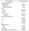
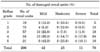
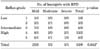
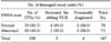
Reflux was unilateral in 88 patients and bilateral in 59, comprising 206 refluxing ureters. VUR was grade I in 19, grade II in 75, grade III in 57, grade IV in 34, and grade V in 21 renal units. An abnormal renal scan was found in 78 (37.7%) renal units (Table 1). An abnormal renal scan was mild in 42 (53.8%), moderate in 25 (33.3%), and severe in 11 (12.9%) renal units. Table 2 shows the abnormal renal scans according to reflux grade. Of the 21 grade V refluxing renal units, 18 (85.7%) showed an abnormal renal scan. By contrast, only 18 (19.1%) of the 94 grade I and II refluxing units showed an abnormal renal scan. The incidence of VUR was significantly higher in males in group 1 (p<0.01) and in females in group 2 (p<0.01). There was no significant difference in the number of abnormal renal scans between males and females in either group (Table 3). The incidence of an abnormal renal scan was significantly higher in infants in group 1 with intermediate and high-grade VUR than in infants with low-grade VUR (p=0.042) (Table 4). The incidence of intermediate and high-grade VUR was not significantly different in group 2. In both groups, there was no significant difference in the number of abnormal renal scans between males and females (p>0.05) (Table 4). Of the five infants screened for sibling VUR, two were boys and three were girls. One infant (20.0%) without a history of UTI had mild renal scarring. On the other hand, 68 of the 138 patients (49.3%) who presented with UTI showed an abnormal renal scan. The incidence of RPD was lower in infants without a history of UTI than in those who presented with UTI. Two of the four infants (50.0%) investigated owing to prenatally diagnosed hydronephrosis had evidence of congenital renal dysplasia without a prior UTI (Table 5).
DISCUSSION
The population of our study consisted of 91 boys (63.0%) and 56 girls (37.0%) with VUR. Group 1 consisted of 75 boys (75.8%) and 24 girls (24.2%). When analyzed on the basis of renal units, the incidence of VUR in group 1 was significantly higher in males than in females (p<0.05). This is in accordance with most other studies, in which most infants with VUR were male [10,11]. Considering that most of our patients (93.9%) had UTI, the dominance of males may be due to a characteristic of this Korean population in which neonatal circumcision is not a common procedure. Compared with group 1, the incidence of VUR was significantly higher in females in group 2 (p<0.05). This is also in accordance with most other studies, in which females represented the major portion of children with VUR. The dominance of females may be due to a characteristic of this population in which UTIs are more common in females in this age period. When we analyzed the incidence of abnormal renal scans by reflux grade, abnormal renal scans were found more often in patients with high-grade reflux, especially in group 1 (p<0.05). This is in accordance with most other studies in infants, in which renal scars are more frequently found in more severe reflux. But this was not the case in the children in our study. Considering that children could also have more severe renal damage after acquired inflammatory reactions in high-grade reflux, they could have more renal scars as well. This difference may be due to the fact that, compared with infants, children can be treated with antibiotics more promptly. This may also be due to the small number in this group in our cohort study and thus imply that further study may be needed in the future.
The report regarding an association of male sex with renal parenchymal damage has been increasingly reported [12,13]. Yeung et al investigated 155 infants with prenatal hydronephrosis who were diagnosed with VUR before the age of 7 months [12]. They found generalized kidney damage in 28% of refluxing units in males and only a 5% incidence in females. Mohanan et al also investigated 549 consecutive infants with VUR and reported that renal scarring was found slightly more often in male than in female infants (28% vs. 25%), and moderate to severe scarring was significantly higher in male than in female infants (73% vs. 27%, p<0.02) [14]. Compared with these studies, our study showed no significant difference in the incidence of an abnormal scan between male and female infants. This may be because, in contrast with other studies in which primarily high-grade VUR in infants was diagnosed after a prenatal diagnosis [12], 93.9% of the patients in our study presented with febrile UTIs. The renal scars detected in these patients may in general be attributable to preceding UTIs. In addition, the size of our study, especially the number of infants with high-grade VUR, was too small to compare statistically. These factors could have affected our findings. It is well recognized that many male infants with high-grade VUR have congenital damage, which currently cannot be prevented. However, it is mandatory to identify VUR early in these infants to prevent exposure to UTIs and avoid the possible progression of renal damage. Whether it is congenital damage or infection-related damage, these infants need early identification and protection from further damage due to ongoing high-grade reflux and/or UTIs. Familial VUR has been described by several investigators and a 27% to 51% prevalence in siblings of children with VUR has been reported [15,16]. In the current study, although the number is too small for comparison, the prevalence of renal damage in infants presenting with UTIs was 49.3%, whereas it was 20.0% in asymptomatic infant siblings with VUR. Significant variability exists in the outcome of acute pyelonephritis following UTIs in VUR in childhood, because some children go on to have permanent renal scarring, whereas others do not. Recently, increasing evidence has suggested that interindividual variation in the susceptibility to renal parenchymal damage may have a genetic basis [17,18]. Improved understanding of the role of genetic factors in the etiology of reflux nephropathy may lead to innovative interventions to prevent parenchymal damage.
CONCLUSIONS
Our data showed that VUR in infants was significantly higher in males than in females, whereas VUR in children was significantly higher in females. This may be due to a characteristic of the population studied, in which neonatal circumcision is not a common procedure in infants and UTIs are more common in female children. Our data also showed that the incidence of an abnormal renal scan was significantly higher in infants with high-grade VUR. Further study may be needed to identify gender differences in renal parenchymal damage in infants with high-grade reflux.

 XML Download