

 PDF
PDF ePub
ePub Citation
 Print
Print


INTRODUCTION
In donor nephrectomy, it is important to understand the exact anatomy of the blood vessels during minimally invasive surgery to decrease hospital stay and blood loss by reducing complications and to accelerate recovery [1,2]. In the past, preoperative evaluation included intravenous pyelography (IVP), angiography, and abdominal sonography [3,4]. With the introduction of muti-detector computed tomography (MDCT) [5], those methods have been replaced by computed tomography (CT) angiography. CT angiography is less invasive than conventional angiography and has the advantage of allowing the assessment of not only the main vessel (renal vein, renal artery) and ureter structure but also renal cystic diseases, renal parenchymal lesions, tiny stones, and hilar vessels (lumbar vein, adrenal vein, gonadal vein) with one test [6-8].
Previous studies have examined the accuracy of CT angiography for determining vessel structures. The accuracy for the main vessels was reported to be 90% to 100% and that for the minor vessels was 75% to 100% [9-12]. One reason for the range in accuracy may be that different CT protocols were used and the protocols used to assess vessel structures during surgery were not consistent. In Korea, similarly, studies have been conducted comparing CT angiography with conventional angiography [12]. Nonetheless, a large-scale prospective study applying 64-channel MDCT has not been conducted.
Therefore, in our study, we prospectively analyzed the accuracy of vessel structures obtained by 64-channel MDCT angiography compared with the actual vessel structure observed during surgery. Efforts were made to evaluate the validation of the hilar vessels, including the right and left main vessel.
MATERIALS AND METHODS
In 335 patients who underwent donor nephrectomy from July 2007 to August 2010, the preoperative evaluation and the postoperative written donor protocol were evaluated prospectively. Before surgery, CT angiography was performed on all patients. Among them, 1 patient who underwent magnetic resonance imaging and 2 patients who were analyzed at another hospital applying a protocol different from the one used in our hospital were excluded. In addition, 94 patients for whom the information recorded in the donor protocol was not sufficient to compare with CT (41 patients) or for whom information was recorded inappropriately (54 patients), such as the loss of the donor protocol, were excluded. The study received institutional review board approval and ethics committee approval.
Among 335 patients, 238 patients satisfied the above criteria. In these 238 patients, the ipsilateral artery, the number of veins, the association with the run of the hilar vessel, and other vascular anomalies recorded during the presurgical CT angiography and the ipsilateral artery, the number of veins (with or without the travels of the hilar vessel), and other vascular anomalies recorded in the prepared donor protocol were summarized.
1. CT protocols
All CT examinations were performed by use of a standardized examination protocol with a multislice 64 detector row helical CT scanner (Lightspeed, GE Medical Systems, Milwaukee, WI, USA). Scanning was initiated with a scout image covering the abdomen. A precontrast image was with 2.5 mm slices; the table rotation time was 0.5 sec with 120 kV and 100 mAs. The arterial phase was obtained 12 s and the venous phase 60 s after the initiation of the contrast bolus. The arterial phase included a volume covering the diaphragm to the pelvis. After acquiring the image, the arterial phase and venous phase images were reconstructed by axial 3 mm and 1 mm images and coronal 3 mm images. Also, a 3D reconstruction image was acquired with 0.625 mm slices. All CT angiographies were read by one radiologist before donor nephrectomy. During the evaluation, the main vessel structure of the right and left side as well as the travel of the hilar vein were described.
2. Donor protocols
All surgeries were performed by 2 surgeons applying video-assisted minilaparotomy surgery (VAMS) techniques [13]. After surgery, the surgeons themselves wrote the protocol. In the protocol, presurgical CT findings, the main vessels actually observed during surgery, the hilar veins, and vascular anomalies were described.
The protocol was prepared prospectively from July 2007, and the age of the patient, sex, operation date, past history, preoperative creatinine level, and creatinine clearance were recorded before surgery and immediately after surgery. In addition, renal vascular structure, special findings observed during surgery, problems occurring during surgery, renal vascular anomalies, and ischemic time were recorded.
The variables tested were compared among the groups by use of the chi-square test, and a p-value of <0.05 was considered significant. Statistical analysis was performed with SPSS ver. 18.0 (SPSS Inc., Chicago, IL, USA).
RESULTS
Of the 238 patients, 101 patients were male and 137 patients were female. Nephrectomy was performed on the left side in 199 patients and on the right side in 39 patients. The mean age of the patients was 39.16 years (range, 17-64 years). Of the two surgeons, one surgeon operated on 67 patients, and the other surgeon operated on 171 patients.
For 238 patients, the results of the main vessel read by CT were compared with the surgical findings. In 199 patients who underwent surgery on the left side, the surgical findings of the drainage of the left lumbar vein detected by CT were evaluated.
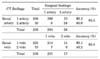
The CT findings and surgical outcomes for the artery and the vein are summarized in Tables 1, 2, 3. The artery and the vein showed 93.3% and 92.4% accuracy, respectively.
For the artery, the accuracy was 95.5% on the left side and 82.1% on the right side, respectively. Regarding vein structure, the accuracy was 93.3% on the left side and 87.2% on the right side, respectively. The accuracy was analyzed by chi-square test. The accuracy of the right side was relatively lower than that of the left side; however, this difference was not statistically significant (artery: p=0.124; vein: p=0.174).
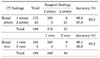
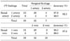
In 199 patients who underwent surgery on the left side, the lumbar vein was recorded by CT and was compared with the actual travel of the lumbar vein as shown in the surgical findings. These results are summarized in Table 4. The overall accuracy of CT was shown to be 84.9%, and accuracy in the group drained to the renal vein was significantly different from that in the group drained to the IVC (p<0.01).
Other points that could be assessed during surgery were that among 199 patients, the left adrenal vein was drained to the renal vein, and the left gonadal vein of 195 patients out of 199 patients was drained to the renal vein.
Among 39 patients who underwent surgery in the right kidney, the right adrenal vein was drained to the renal vein in 4 patients, and the right lumbar vein was drained to the renal vein in 3 patients.
Double IVC was confirmed in 3 patients during surgery, and early bifurcation was detected in 22 patients during surgery.
DISCUSSION
Regarding the accuracy of the vessel anatomy as recorded by MDCT, the accuracy of the renal vascular anatomy was reported to be 90% to 100% and that of minor venous abnormalities was reported to be 75% to 100% in several studies. Nonetheless, in most studies, the number of subjects was limited in many cases. In addition, cases in which 64-channel MDCT was applied were rare. In addition, in many studies with high accuracy, CT reading was performed retrospectively, and thus the accuracy measured may have been high [14].
The advantages of our study were that the results of presurgical 64-channel MDCT and the surgical findings prepared immediately after surgery were analyzed, and thus the data were analyzed prospectively, and the study was conducted in a relatively large number of patients (n=238).
Raman et al reported in 2006 that the sensitivity of MDCT data for detection of the renal artery was 65/66 (98.5%) and that for the vein was 61/63 (98%) [15]. In Korea, in 2002, Roh et al reported that the accuracy of CT for the artery and the vein was 91% and 95%, respectively [12].
Our study was conducted on a large patient group, 238 patients, and the accuracy of the MDCT for the artery and the vein was 93.3% and 92.4%, which was similar to previous studies. In addition, the accuracy of both arteries and veins was analyzed, and the accuracy of the right side was relatively lower than that the left side, although not significantly so.
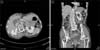
In minimally invasive surgery, because of the limited field of view, assessment of the main vessels and the hilar vessels before surgery can be considered to be important [16]. There are examples, Fig. 1 and Fig. 2 shows actual surgical field in operation. Case of Fig. 1, MDCT finding was exact to actual anatomy. Fig. 3 is example of actual renal vescular structure was different from MDCT findings. As CT becomes more accurate, not only the main vessels but also the hilar vessels can be assessed. Nevertheless, regarding the hilar vessels, reports of accuracy were limited in several studies, and the patient group was limited in many cases. Lewis et al reported the accuracy of the hilar veins in 2004. The accuracy for the gonadal vein was 33/37 (83%), that for the adrenal vein was 27/33 (83%), and that for the lumbar vein was 13/20 (75%) [17]. Bhatti et al reported that the accuracy for the gonadal veins was 74% and that for the lumbar veins was 81% [18].
In our study, we assessed the accuracy of MDCT for the lumbar vein in 199 cases who underwent left donor nephrectomy. The accuracy was 84.9%, which is relatively higher than in previous studies. However, in 66 cases in which the lumbar vein was drained to the IVC, the accuracy was observed to be 54.1% (p<0.01). Thus, it appears that attention should be paid to the interpretation of the CT results for the lumbar vein.
In many cases, of the two sides, the side with the less complex vascular structure is selected as the surgical side. Thus, it is thought that the measured vascular accuracy may be higher than actual values, and studies of this are required in the future. In addition, in our hospital, studies on the accuracy for the lumbar vein were conducted; nonetheless, it appears that additional studies of accuracy for the gonadal vein and the adrenal vein are required.
In another study conducted on 615 cases of donor nephrectomy performed at our hospital from 2003 to 2009, the overall complication rate was 35 cases (5.9%) [19]. Among the 35 cases, complications caused by the upper pole renal artery undetected by CT occurred in 14 cases, which were most prevalent. Complications caused by a missing renal vein occurred in 4 cases, and complications in the adrenal vein and lumbar vein occurred in 1 case each.
When we examined 24 cases of complications that developed in 168 cases during the same period as our study (July 2007 to December 2009), in 21 cases whose main vessels were reported erroneously, complications developed in 4 cases. Nonetheless, statistical differences were not shown (19%, p=0.27).
In regard to the main vessels, the accuracy of 64-channel MDCT angiography was 93.3% and 92.4% on both sides of the artery and vein. The accuracy of the right side artery was 82.1%, which was relatively lower than that of the left side of 95.5%. Nonetheless, statistical significance was not detected. Raman et al and Janschek et al reported that whereas the frequency of renal venous variants was higher for the left side than for the right side, the frequency of major venous anomalies was higher for the right side kidney [15,20]. The length of the vascular pedicle on the right side kidney is generally shorter than on the left side, which may have caused the relatively lower accuracy for the right side kidney.
The accuracy of the left lumbar vein was 84.9%, and when the lumbar vein was drained to the IVC, the accuracy was as low as 54% (p<0.01). Thus, it may be necessary to pay attention to the interpretation of the lumbar vein.
CONCLUSIONS
MDCT angiography is important for understanding the exact anatomy of blood vessels before surgery, especially during minimally invasive surgery. We showed 64-channel MDCT to have high accuracy for the main vessel and hilar vessels. However, it may be necessary to pay attention to the interpretation of the lumbar vein.


 XML Download