

 PDF
PDF ePub
ePub Citation
 Print
Print


Liposarcoma is the most common tumor type of retroperitoneal soft tissue sarcomas [1]. Retroperitoneal liposarcomas are difficult to detect early because the symptoms of these tumors appear late and are nonspecific; the tumors can thus grow slowly in the retroperitoneal space, reaching a considerable size before being diagnosed [2]. Case reports of retroperitoneal liposarcomas with a maximal diameter >15 cm have often been made. Regardless of size, the treatment of choice for a huge retroperitoneal sarcoma is surgery. Complete resection of the tumor and a favorable histopathologic grade are positively associated with long-term disease-free survival. Compared with other retroperitoneal sarcoma subtypes, liposarcoma of this area shows a relatively good prognosis. Effects of chemotherapy and radiation therapy have also been reported. In some cases, these treatments have been reported to be effective as an adjuvant therapy, but controversy over this point still exists. We experienced two cases of giant retroperitoneal liposarcomas treated at our medical institution. Here we discuss the histopathologic findings and surgical outcomes of these cases. We report our cases with a review of past studies.
CASE REPORTS
Case report 1
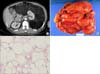
An 82-year-old man presented at our institution with a chief complaint of an abdominal mass and associated abdominal discomfort. He had first noticed it 4 years before the consultation, and it was revealed as a lipomatous retroperitoneal tumor by computed tomography (CT) performed at a local clinic. The clinic referred the patient to us because the serial follow-up imaging study showed that the tumor had been growing. During the physical examination, we were able to palpate the large abdominal mass. We found no other specific abnormalities associated with it. Suspecting it to be a liposarcoma, we performed an abdominal CT scan and saw a huge abdominal mass lesion composed mainly of lipomatous tissues (Fig. 1A). All laboratory data were within normal ranges. We made a preoperative diagnosis of a retroperitoneal liposarcoma, and the patient was explored via laparotomy.
Intraoperatively, there was a mass occupying the abdominopelvic cavity. Because the lesion involved the left kidney and adrenal gland, we performed a tumor resection en bloc with the left kidney and the adrenal gland. The surgical specimen measured 30×30×8 cm and weighed 3,600 g (Fig. 1B). The cut surface of the mass was multilobular and almost uniformly yellow. The histopathological diagnosis was a well-differentiated liposarcoma originating from the retroperitoneum (Fig. 1C).
No evidence of recurrence or metastasis was noted on the follow-up CT scans conducted 1.5 years after the operation.
Case report 2
A 64-year-old man presented with a known huge abdominal mass. An ultrasound-guided biopsy had already been performed at another hospital, and a retroperitoneal liposarcoma was reported. Because the patient had complained of mild dyspnea and bowel habit changes, he was sent to our institution for further evaluation and treatment. An abdominal CT scan was performed for re-examination, demonstrating an 11.5×9×17.5 cm sized fatty mass with an enhanced solid portion located in the left renal subcapsular area (Fig. 2A). We assumed it was a retroperitoneal lipomatous tumor and decided on surgical exploration. The surgery revealed a huge retroperitoneal mass, measuring 18 cm in maximal diameter. Because the tumor grossly encased the left kidney, we also performed a nephrectomy for complete resection of the tumor. The final pathological report confirmed a well-differentiated liposarcoma, and the resection margins were tumor-free (Fig. 2B, C). The postoperative course was not significant, and the follow-up imaging study showed no evidence of tumor recurrence after 1.5 years.
DISCUSSION
The annual incidence of sarcoma is relatively low, composing approximately 0.8% of all newly diagnosed malignancies. Of these sarcomas, liposarcoma accounts for approximately 20%, and about 13% of them originate from the retroperitoneum. Leiomyosarcoma and pleomorphic subtypes are also common cases of retroperitoneal sarcomas. Retroperitoneal liposarcomas are then further classified by their histologic grades of cellular differentiation.
In most patients, definite symptoms are lacking, and the diagnosis is routinely made on the basis of CT scans of the abdomen and pelvis. Abdominal organ involvement and local metastasis are relatively common [3].
Although controversy exists, adjuvant or neoadjuvant radiotherapy or chemotherapy do not have any survival benefits in retroperitoneal liposarcoma. Only surgical intervention has been shown to improve overall survival, and the most important variable in outcome is the ability to completely resect the tumor, which often requires removal of the adjacent organs. In a previous study, the median survival of patients who underwent complete resection was 103 months. Conversely, the median survival of patients undergoing incomplete resection was 18 months, which was no different from performing no resection at all [4].
Histopathologic grades of differentiation and tumor subtype also affect overall survival rates. Of the subtypes, the 5-year survival rate of well-differentiated liposarcoma reaches 90%. By contrast, the survival rate of pleomorphic subtypes has been reported to be 50%. In addition, the survival rate has been reported to be 75% in de-differentiated cases and 60% in cases of the myxoid/round cell subtype [1,5].
Mortality associated with retroperitoneal sarcoma is usually due to its local recurrence. In a prospective study of 500 patients with retroperitoneal sarcoma, the median survival was significantly lower in a group with local recurrence than in a group with only primary disease [4]. Although the rate of local recurrence was higher than for other sarcomas, meaningful long-term survival could be achieved with multiple re-operations for recurrent disease [6].
Because there are few clinical findings and specific laboratory abnormalities, these tumors may grow to a large size without any symptoms. The literature shows that about 20% of retroperitoneal sarcomas are>10 cm at the time of diagnosis [4,7]. For example, Yol et al reported a retroperitoneal tumor weighing 42 kg [8].
The size of the tumor itself is not associated with surgical resectability and consequently does not affect survival. In a certain group of patients who underwent complete resection, however, a size>10 cm and a high histologic grade could predict poorer treatment outcome.
As in the presenting cases, the tumors are usually large at the time of detection, which corresponds with the difficulties of achieving complete surgical excision [9]. However, we could demonstrate complete tumor resection with a negative margin, whereas the reported negative microscopic margin rate in general was 74% in a past study [4]. That study also described the perioperative (30-day) mortality rate (4%), related to complications such as bleeding, sepsis, and myocardial infarction. Neither of our patients had any serious perioperative or postoperative events. After 1.5 years, the patients underwent follow-up CT, and no tumor recurrence was detected. From this point, combined with the fact that we achieved negative surgical margins and the tumors were of a good histopathologic grade and subtype, we predict a low rate of local recurrence and good overall survival.
Complete resection is thus the most important component of treatment, regardless of tumor size or adjacent organ involvement. The resectability of the tumor and histopathologic grades are associated with disease prognosis, and both factors are hard to elucidate before undergoing surgical intervention. Resections of contiguous organs are common and include en bloc resection of retroperitoneal organs, including the kidney, adrenal gland, pancreas, spleen, and even vascular resection (inferior vena cava) if indicated [4].
Several retrospective studies have suggested that postoperative radiotherapy may yield better outcomes than surgery alone, although other similarly designed studies revealed no advantage [10]. Randomized large-sized group trials are required to determine the benefit of adjuvant radiotherapy after surgery.

 XML Download