

 PDF
PDF ePub
ePub Citation
 Print
Print


INTRODUCTION
Selective serotonin reuptake inhibitors (SSRIs) have previously been used as a treatment for premature ejaculation. The use of SSRIs for the treatment of premature ejaculation began after it was discovered that treatment of depression in males with SSRIs resulted in a side effect of delayed ejaculation [1]. Sertraline is an SSRI, and its treatment efficacy for premature ejaculation has also been reported [1].
Current-source analysis of electroencephalography (EEG) is an electrographic and functional imaging method that is used to find the current source of neuronal activity of the brain. It involves a computational and mathematical algorithm that analyzes digital EEG data. The discrete model of current-source analysis provides information about location, strength, and orientation of current-source dipoles. The distributed model is a method used to find the location and extent of the current source distribution. Low-resolution electromagnetic tomography (LORETA) is a distributed model of current-source analysis [2].
Studies of neurocognitive function using EEG data have focused on the frequency band above 20 Hz. It has been shown in human EEG studies that cortical high-frequency activity is topically enhanced in various cognitive processes [3]. Complex cognitive tasks ignite neuronal cell assemblies and activate loops of neuronal networks. In the case of language processing, a loop between Broca's and Wernicke's regions is activated [4]. The neuronal oscillations have a circulation frequency of 25 to 30 Hz. In contrast, higher-frequency oscillatory cortical activity, in the range of 40 to 60 Hz, is observed in less widely dispersed cell assemblies [5]. The highest frequency band that can be analyzed by LORETA is the high beta frequency band (22-30 Hz). The analysis of this frequency band may be helpful for evaluating neurocognitive functions in various situations.
Little is known about the cerebral control mechanisms of ejaculation. According to our previous study using EEG recordings from patients with premature ejaculation (PE) [6], we observed decreased neuronal activity in the precentral gyrus and the insula of the right cerebral hemisphere and in both superior parietal lobules during sexual arousal induced by erotic videos. In addition, the neuronal activity of PE patients decreased in the right parahippocampal gyrus and in the left middle temporal gyrus compared with normal controls. These findings suggest that the inhibitory control of ejaculation in the central nervous system may be impaired in PE patients. The purpose of this study was to examine the changes in the brain current-source density of the high beta frequency band (22-30 Hz) induced by sertraline administration and erotic stimuli in healthy adult males.
Go to : 
MATERIALS AND METHODS
1. Subjects
Before the study, the participants were interviewed to assess their motivation for participating in the study and to exclude anyone with a major medical disorder or history of drug abuse, psychiatric disorder, or erectile dysfunction. All subjects received a detailed explanation of the study design, and written informed consent was obtained from all subjects. All 11 subjects (mean age, 25.90±1.22 years) enrolled in this study. All participants were right-handed as determined by using the Chapman-Chapman handedness scale [7].
2. Electroencephalography
The EEGs were recorded in a comfortable, closed room. A male technician attended each patient to monitor the EEG recording state and to mark each of the time points for the experiments. Based on the international 10-20 system, all EEGs were recorded by using 25 channels (Fp1, Fp2, F3, F4, C3, C4, P3, P4, O1, O2, F7, F8, T3, T4, T5, T6, Fz, Cz, Pz, F9, F10, T9, T10, P9 and P10) at a sampling rate of 400 Hz. Scalp EEGs were conducted twice for each volunteer, once before the intake of 50 mg sertraline and again 4 hours thereafter. These time points were chosen because it is well known that the mean peak plasma concentrations (Cmax) of sertraline occur 4 to 6 hours after dosing. The EEGs included four segments recorded sequentially while the participant was resting, watching a music video, resting, and watching an erotic video for 3 minutes, 5 minutes, 3 minutes, and 5 minutes, respectively [8]. The visual angles of the monitor used to display the visual stimuli were 30.5° horizontal, 23℃ vertical, and 38℃ diagonal.
3. Self-questionnaire on sexual excitement level
After completion of the EEGs, the participants completed a self-questionnaire about the level of sexual excitement felt while watching the erotic videos. Sexual excitement without any erection was 0, and an erection at any level was recorded as 1. All eleven participants recorded their excitement level as 1.
4. Current-source analysis
After recomputing to the average reference montage, five 5-s artifact-free segments were selected from the sessions of erotic video and music video in each EEG (filter: 1.6-70 Hz). We used the data review and processing module in the brain electrical source analysis (BESA v. 5.1, MEGIS, Grafelfing, Germany) to obtain the data. These segments were then subjected to a cross-spectral analysis. Frequency-domain analysis in the 22 to 30 Hz frequency band was applied to the selected 5-s artifact-free segments. LORETA-KEY (KEY Institute for Brain-Mind Research, Zurich, Switzerland) was used to calculate the intracerebral current density in a cross-spectral analysis of the segments. LORETA powers were calculated by using the software [2,9].
Our version of LORETA used a three-shell spherical head model registered to the Talairach human brain atlas and available as digitized magnetic resonance images from the Brain Imaging Center of the Montreal Neurologic Institute [10]. The registration between the spherical and Talairach head geometries used the realistic EEG electrode coordinates reported by Towle et al [11]. The LORETA solution space was restricted to the cortical gray matter and hippocampus in the Talairach atlas, as defined by the corresponding digitized probability atlas available from the Brain Imaging Center of the Montreal Neurologic Institute. A total of 2,394 voxels was produced at 7 mm spatial resolution under this neuroanatomical constraint [2].
5. Statistical analysis
In each patient, five segments of 5-s epochs were selected from the EEG recordings made during the watching of music video and erotic video excerpts before and 4 hours after the intake of sertraline. Thus, a total of 110 EEG data epochs (11 subjects x10 epochs) were obtained under each condition. Paired-sample t-tests were computed for the log-transformed LORETA power at each voxel in the high-beta frequency band (22-30 Hz) to evaluate differences between the erotic and music video sessions before and 4 hours after sertraline intake. In all analyses, statistical significance was set at p<0.05. These voxel-by-voxel t-values were displayed as statistical nonparametric maps (SnPMs). Talairach coordinates, and anatomical locations of the areas with significant changes in current-source density induced by the erotic stimulation were obtained by using the SnPM before and 4 hours after sertraline intake. The SnPMs between the music video sessions 4 hours after the intake and those sessions before the intake, and between the erotic video sessions 4 hours after the intake and those sessions before the intake were also taken (p<0.05).
Go to :
RESULTS
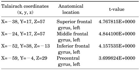
In the SnPMs comparing the erotic video sessions with the music video sessions before sertraline intake, the current-source density of the high beta frequency band decreased in the middle frontal gyrus, the precentral gyrus, the postcentral gyrus, and the supramarginal gyrus of the left cerebral hemisphere (Table 1, Fig. 1). Four hours after sertraline intake, no differences in current-source distribution between erotic and music video sessions were observed. In the SnPMs comparing the erotic video sessions 4 hours after the intake with those sessions before sertraline intake, the current-source density of the high beta frequency band increased in the superior, middle, and inferior frontal gyri and the precentral gyrus of the left cerebral hemisphere (Table 2, Fig. 2). There was no change in the high beta frequency band in the SnPMs comparing the music video sessions 4 hours after the intake with those sessions before sertraline intake.
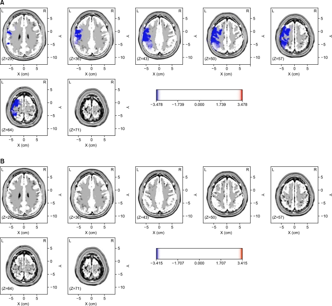 | FIG. 1Statistical nonparametric maps (SnPMs) comparing the current-source distribution of the high beta frequency band (22-30 Hz) of erotic video sessions and those of music video sessions in healthy, right-handed young males. The current-source density decreased in the postcentral gyrus, the precentral gyrus, the supramarginal gyrus, and the middle frontal gyrus of the left cerebral hemisphere under audiovisual erotic stimulation before sertraline intake (A). The changes disappeared in the SnPMs 4 hours after sertraline intake (B).
|
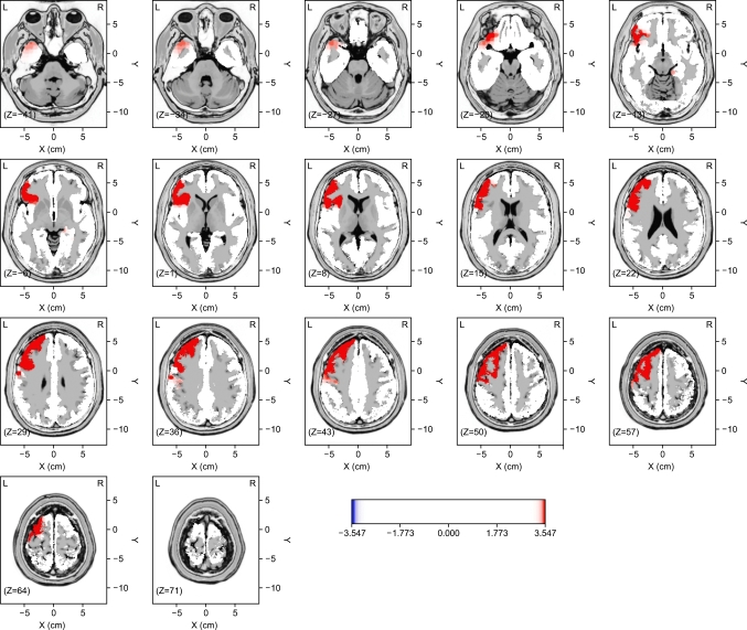 | FIG. 2Statistical nonparametric maps (SnPMs) comparing the erotic video sessions 4 hours after sertraline intake and those sessions before sertraline intake. The current-source density of the high beta frequency band increased in the superior, middle, and inferior frontal gyri and the precentral gyrus of the left cerebral hemisphere.
|

Go to :
DISCUSSION
It is known that the serotonin pathway plays a role in the mechanism of ejaculation in males. In the central nervous system, the medial preoptic area, nucleus paragigantocellularis, stria terminalis, amygdala, and thalamus may be the anatomical locations associated with this mechanism [12-14]. Serotonergic pathways descending from the nucleus paragigantocellularis to the lumbosacral motor nuclei tonically inhibit ejaculation. When the signal from the medial preoptic area inhibits the nucleus paragigantocellularis, the motor nuclei in the lumbosacral spinal area are released from the tonic inhibition of the nucleus paragigantocellularis, which allows ejaculation [13].
In our previous study, we found that the neuronal activity of PE patients decreased in the precentral gyrus and the insula of the right cerebral hemisphere and in both of the superior parietal lobules during sexual arousal induced by erotic videos [6]. In addition, the activity decreased in the right parahippocampal gyrus and in the left middle temporal gyrus compared with the normal controls. These results suggested that the debasement of inhibitory control in the central nervous system located above the medial preoptic area may be a cause of premature ejaculation.
When SSRIs are administered to patients for the treatment of depression, the brain activity measured by functional magnetic resonance imaging (fMRI) decreases in the cingulate gyrus and the caudate nucleus [15]. Positron emission tomography (PET) and fMRI are methods used to evaluate neural activity indirectly by observing local cerebral blood flow or metabolism without observing neuronal activity directly. In contrast, the current-source analysis of EEGs observes the electric activity of neurons directly. Therefore, results from the current-source analysis of EEGs may differ from the results of studies using PET or fMRI.
We recorded EEGs before and 4 hours after sertraline intake in healthy, right-handed young males under erotic stimulation using sexual video excerpts and analyzed the current source of the high beta frequency band by using a distributed model. Erotic stimuli decreased the current source density of the high beta frequency band in the middle frontal gyrus, the precentral gyrus, the postcentral gyrus, and the supramarginal gyrus of the left cerebral hemisphere in the baseline EEGs (p<0.05) (Table 1, Fig. 1). These changes were not observed 4 hours after sertraline was administered. In addition, we also observed that sertraline increased the current-source density in the superior, middle, and inferior frontal gyri and the precentral gyrus of the left cerebral hemisphere (p<0.05) (Table 2, Fig 2). According to these results, the erotic stimuli may trigger a functional debasement of inhibitory controls in the middle frontal gyrus, the postcentral gyrus, the precentral gyrus, and the supramarginal gyrus of the left cerebral hemisphere, which may lead to the sexually excited state. It is also possible that sertraline may suppress these cerebral changes and decrease sexual excitement. In addition, sertraline may reduce the excessive sexual excitement in the brain, prolong the duration of an erection, and suppress ejaculation.
The prefrontal cortex is known to be a crucial area for cognitive control [16], and the dorsolateral prefrontal cortex (DLPFC) is involved in self-control [17]. In a study observing the reaction of the cerebral cortex to meals in right-handed females, the decreased activity of the left DLPFC was associated with poor control of appetite and resulting obesity [18]. In another study investigating the association between the stop-signal task and response inhibition, the precentral gyrus of the left cerebral hemisphere was activated during response inhibition, and this activation was associated with the stop-signal reaction time [19]. Our results are similar to these reports in showing that the activity of the middle frontal gyrus and the precentral gyrus of the left cerebral hemisphere decreased during sexual arousal in right-handed healthy young males. These changes recovered after administration of sertraline. Taken together, these findings suggest that the debasement of inhibitory control by erotic stimuli in the middle frontal gyrus and the precentral gyrus of the left cerebral hemisphere may lead healthy young males to be excited sexually. These results also suggest that inhibitory control may be recovered by the effects of sertraline.
The supramarginal gyrus of the left cerebral hemisphere is responsible for the detection of changes in phonological units [20]. The postcentral gyrus is a somatosensory area, and lesions in the left postcentral gyrus induce sensory changes in the contralateral face and body [21]. Even though it is difficult to interpret the changes induced by erotic stimuli in those areas, the aforementioned structures may also be a part of the cerebral inhibitory center that controls sexual excitement in males.
We used the small number of 25 available channels, which could have resulted in some localization errors, particularly in the basal aspects of the brain [22]. The errors associated with using a spherical model with the small number of electrodes range from 10 to 20mm [22-24]. The localization accuracy for LORETA has been shown to increase using 25 to 89 electrodes and to plateau thereafter [25]. However, to localize the areas of increased activity, the small number of electrodes may be sufficient for the SnPM method of LORETA [26,27]. It has also been observed that current source estimation using LORETA in a three-shell head model is similar when using between 19 and 46 scalp electrodes when the electrodes are evenly distributed. To investigate cognitive function, lobar-level information may provide useful information, which is not the case for the current-source analysis of epilepsy.
The changes in cerebral activity in response to erotic stimuli may differ depending on subject age [28]. If the subjects were older, the results of this study could be different. The results and interpretation of this study may be limited to young males. Another limitation of this study is that we did not assess the objective level of sexual excitement by using penile erection status or other measures. We addressed this limitation by using a self-report questionnaire to evaluate the status of sexual excitement, and all subjects reported that they experienced penile erection with sexual excitement. Even though the repetitive exposures to erotic video could reduce the sexual arousal, the results of this study may still be reliable. Because the refractory period of young adults may be short, the re-exposure to the same erotic video after 4 hours may not reduce the sexual arousal that much. In addition, all attendants also reported the penile erection with sexual arousal in the questionnaires taken after the 2nd EEG sessions.
More advanced studies can be based on the results presented here. Similar research designs could be adapted for future studies including healthy people or patients with sexual dysfunction in a range of age groups. The reliability of information could also be improved in a variety of ways in future studies. We could use more objective monitoring, by measuring, for example, penile tumescence, and could conduct placebo-controlled studies. Such research designs may be useful for pharmacological studies of many other drugs for sexual dysfunction.
Go to :
CONCLUSIONS
The current-source density of the high beta frequency band decreased following sexual arousal in the middle frontal gyrus, the postcentral gyrus, the precentral gyrus, and the supramarginal gyrus of the left cerebral hemisphere in healthy, right-handed young males. The changes in current-source density were not observed 4 hours after sertraline intake. Taken together, these findings indicate that erotic stimuli may decrease inhibitory control against sexual arousal in the middle frontal gyrus, the postcentral gyrus, the precentral gyrus, and the supramarginal gyrus of the left cerebral hemisphere. Sertraline may influence cortical activity and recover the debasement of inhibitory control at higher levels of the brain.
Go to :
 XML Download