

 PDF
PDF ePub
ePub Citation
 Print
Print


INTRODUCTION
Urolithiasis is a common disorder that affects approximately 3-5% of the population and is usually associated with metabolic abnormalities [1-3]. Although both intrinsic and environmental factors contribute to urinary stone formation, the exact cause is not fully understood [2-7]. More precise knowledge about the etiology of urolithiasis may contribute to improvements in disease management and prevention.
Hypercalciuria has been considered to be the most common abnormality identified in calcium stone formers (SFs) [2,3]. Recent studies have suggested that a defect in phosphate balance, such as maximal reabsorption of phosphate by the glomerular filtration rate (TmP/GFR), is a significant underlying cause of calcium urolithiasis [8-11]. Reduced renal phosphate reabsorption leads to hypophosphatemia that, in turn, increases 1,25(OH)2-vitamin D synthesis and enhances the intestinal absorption of phosphate and calcium, resulting in hypercalciuria [12-14]. Additionally, mutations in the genes that encode two renal sodium phosphate co-transporters, NPT2a and NPT2c, have been identified in patients with hyperphosphaturia [15,16]. Inactivation of the gene that encodes NPT2a results in increased excretion of phosphate and calcium and bone demineralization [17]. Thus, there has been increasing interest in the role of renal phosphate handling and the associated hyperphosphaturia in stone formation.
Identification of modifiable risk factors for urolithiasis may result in new approaches to the treatment and prevention of this disorder. The present study was designed to assess whether phosphaturia relates to urinary metabolic abnormalities and recurrent stone formation.
MATERIALS AND METHODS
1. Subjects and sample collection
A database of calcium SFs who were diagnosed and treated between 1994 and 2008 at our institute was reviewed. The database included stone history, medication, and a metabolic evaluation that included serum chemistry, 24-hour urinary chemistry, and radiographic imaging of the urinary tract. A diagnosis of calcium stone formation was based on an abdominal radiograph and ultrasound examination. The metabolic evaluation was performed at least 6 weeks after the last stone episode. SFs with any of the following conditions were excluded from the analysis: pediatric patients (<16 years), incomplete 24-hour urine collection, impaired renal function (serum creatinine >1.5 mg/dl), staghorn calculi, urinary tract obstruction, malformation of the urological system, hypercalcemia, prior diagnosis of primary hyperparathyroidism, or other systemic diseases that might affect calcium and bone metabolism. Medications that could affect biochemical parameters were discontinued 2 weeks before the 24-hour urine collection. SFs were told to continue their usual diet, and no patient had been placed on a low-calcium diet. Thus, 1,068 consecutive calcium SFs [706 first-time stone formers (FSFs) and 362 recurrent stone formers (RSFs)] with complete metabolic assessments were included in the final analysis. Of these, SFs who had been followed up for more than 36 months or had stones during the follow-up period (median, 46.0 months; range, 5-151 months) were eligible for analysis of stone recurrence.
After informed consent was obtained, 106 healthy volunteers (mean age: 42.2±12.8 years [range, 17 to 72 years]; 67% male [69/106]; 34.7% female [37/106]) who had no evidence of stone disease as confirmed by abdominal radiograph and ultrasound examination, unimpaired renal function, no history of urological disease, and no medications were enrolled as controls.
2. Measurement and definition of parameters
All measurements were made by routine methods. In the normal controls, 95% of the subjects had a urinary phosphate excretion of less than 800 mg/day. Because there is no universally accepted definition for hyperphosphaturia, any value for phosphaturia that was higher than 95% of the control values was defined as indicative of hyperphosphaturia [8,9]. Urinary metabolic abnormalities were classified into five categories as follows: low urine volume (urine volume less than 2,000 ml hypercalciuria (calcium greater than 300 mg in males and 250 mg in females), hyperoxaluria (oxalate greater than 45 mg), hyperuricosuria (uric acid greater than 800 mg in males and 750 mg in females), and hypocitraturia (citrate less than 320 mg) [3]. Stone recurrence was defined as the passage, extracorporeal removal, endoscopic or surgical removal, or radiographic appearance of stones that were not present on previous examination. The time to stone recurrence was measured from the date the patient was pronounced as being stone free at our institution to the date of stone relapse during the follow-up period or last follow-up.
3. Statistical analyses
To investigate the effect of phosphaturia on urinary metabolites and recurrent stone formation, subjects were classified as SFs with normophosphaturia or as SFs with hyperphosphaturia. Differences in the urine variables between the subgroups were assessed by Student's t-tests. Categorical variables were compared with chi-square tests. The Kaplan-Meier method was used to calculate the time to recurrence, and differences were assessed with log-rank statistics. The prognostic value for the status of phosphaturia on stone recurrence was studied with a Cox proportional hazards regression model. Cases were adjusted for well-known clinical and metabolic prognostic factors for recurrence. All statistical analyses were performed with SPSS 12.0 software (SPSS Inc., Chicago, USA), and p-values<0.05 were considered statistically significant.
RESULTS
1. Baseline characteristics
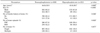
The 1,068 patients were grouped according to whether or not they had hyperphosphaturia (80.1% of the SFs had normophosphaturia [856/1,068]; 19.9% of the SFs had hyperphosphaturia [212/1,068]). The mean age was significantly different between the two groups. Compared with FSFs, the incidence of hyperphosphaturia was more common in RSFs. The baseline characteristics of the SFs are presented in Table 1.
2. Urine parameters
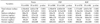
SFs with hyperphosphaturia had greater urinary volume and higher levels of calcium, uric acid, oxalate, and citrate than did SFs with normophosphaturia. The mean urinary pH was lower in SFs with hyperphosphaturia than in SFs with normophosphaturia. Subgroup analyses, grouped by stone episodes, also showed similar results (Table 2).
3. Status of phosphaturia and stone recurrence
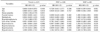
Two hundred forty-seven (23.1%) SFs were eligible for analysis of recurrent stone formation (21.1% of the FSFs [149/706] and 27.1% of the RSFs [98/362]). During the follow-up period, 62.3% (154/247) had recurrent stones (57.0% of the FSFs [85/149] vs. 70.4% of the RSFs [69/98], p=0.034). Kaplan-Meier estimates revealed significant differences in the prevalence of recurrent stone formation between patients with hyperphosphaturia and those with normophosphaturia (log-rank test, p=0.032) (Fig. 1A). Stratified by stone episodes, FSFs with hyperphosphaturia had a shorter time to stone recurrence than did FSFs with normophosphaturia (log-rank test, p=0.010) (Fig. 1B), whereas the time to recurrence was not affected by hyperphosphaturia in the RSFs (log-rank test, p=0.931). Multivariate Cox regression analyses that included known clinical (mean age at stone onset, sex, stone episodes, family history of stones, and stone multiplicity) and metabolic (low urine volume, hypercalciuria, hyperuricosuria, hyperoxaluria, and hypocitraturia) risk factors for stone recurrence revealed that hyperphosphaturia (hazard ratio [HR]: 1.718; 95% confidence interval [CI]: 1.090-2.707; p=0.020) was significantly associated with stone recurrence in SFs. Stratified by stone episodes, hyperphosphaturia (HR: 2.122; 95% CI: 1.100-4.097; p=0.025) was the only independent predictor for stone recurrence in FSFs. In contrast, hyperphosphaturia was not associated with stone recurrence in RSFs (Table 3).
DISCUSSION
Recently, several studies have shown that patients with idiopathic hypercalciuria have phosphaturia or renal phosphate leakage [8,9,12,16]. Renal phosphate leakage and the associated phosphaturia result in increased production of 1,25(OH)2-vitamin D3, which causes increased intestinal phosphate and calcium absorption. The combination of hyperphosphaturia and hypercalciuria from the increased intestinal calcium absorption favors the formation of calcium phosphate complexes that can result in urolithiasis [11]. In a previous study, 19% of SFs had renal phosphate leakage, defined as renal phosphate threshold values lower than 95% of the normal control population, which was associated with high urinary calcium excretion [8]. Similarly, our results showed that the incidence of hyperphosphaturia was 19.9%, which partly supports findings showing that renal phosphate leakage is frequently detected in patients with stone disease.
Studies of phosphate excretion have shown that phosphaturia plays a key role in stone formation [18,19]. Calcium phosphate crystals are injurious to cells of the proximal tubules and the collecting ducts. Calcium phosphate crystals can interact with the renal epithelium, promote sites for crystal attachment, and then either grow into mature calcium phosphate stones or create sites for calcium oxalate crystal formation, allowing calcium oxalate crystals to grow into clinically significant kidney stones [18,19]. In our study with long-term follow-up of patients, hyperphosphaturia significantly affected not only urinary metabolites but also recurrent stone formation. Although the exact biological mechanism is still not known, the results of previous studies provide possible explanations [8,16-22]. Impaired renal phosphate reabsorption increases the risks of nephrolithiasis and bone demineralization [8,9,16,17]. More recently, several reports suggested that the renal phosphate transporters are an important cause of phosphaturia in patients with urolithiasis [17,20-22]. NPT2a is present in the apical membrane of the renal proximal convoluted tubule and is responsible for reabsorption of 80% of the phosphates filtered at the glomerulus [20]. Regardless of the serum phosphate concentration, phosphate is freely filtered across the glomerulus and reabsorbed along the renal tubule, primarily the proximal tubule, through two distinct sodium phosphate transporters that are dependent on the sodium gradient [11]. Thus, inactivation of associated genes results in increased excretion of phosphate and calcium [17]. In addition, the sodium-hydrogen exchanger regulatory factor 1 (NHERF1) can interact with known amino acid sequences at the C-terminal end of NPT2a, which induces alteration of NPT2a targeting [23,24]. Targeted gene deletion of NHERF1 provokes not only increased excretion of phosphate, calcium, and uric acid, but also interstitial deposition of calcium, primarily in the papilla of the kidney, which are prone to the formation of calcium stones, uric acid stones, or both [21]. These findings support our results showing that hyperphosphaturia was significantly associated with increased urinary excretion of calcium and uric acid as well as stone recurrence. However, it is uncertain why hyperphosphaturia was associated with recurrent stone formation only in FSFs. There have been several reports of metabolic differences, such as urinary excretion of citrate, uric acid, and urine volume, between FSFs and RSFs [7,25]. These differences may lead to the inconsistent effect of hyperphosphaturia on recurrent stone formation in FSFs and RSFs. Our data also demonstrated that hyperuricosuria was an independent risk factor predicting recurrent stone formation in RSFs. This finding is supported by earlier reports [7,26].
Uric acid crystals act as a nidus for calcium oxalate stone formation by altering the solubility or precipitation product of calcium and oxalate in solution [22,27]. A recent study also indicated that NHERF1 interacts with the mouse urate transporter 1 (mURAT1) to regulate uric acid transport in the renal proximal tubule and that uric acid excretion is increased in NHERF1-knockout mice [22]. Patients with gout display a significantly reduced tubular reabsorption of phosphate and renal phosphate threshold, which results in hyperphosphaturia [28]. These observations support our findings of a close relationship between hyperphosphaturia and increased urinary excretion of uric acid. However, it is unclear why hyperphosphaturia was closely correlated with other urinary metabolites, such as urine volume, pH, oxalate, and citrate, in our results. These associations may be due to not only various loads of dietary phosphate, but also the existence of currently unidentified mechanisms or interactions. Dietary factors are suggested to be the major sources of urinary phosphate excretion [29]. Accordingly, this may cause the paradoxically increased excretion of citrates and urine volume, which are well-known urinary stone inhibitory factors, in SFs with hyperphosphaturia. Further research is needed to uncover these relations.
In the clinical setting, identification of meaningful, modifiable risk factors is useful for reducing the prevalence of recurrent stone formation. Our results suggest that hyperphosphaturia is an independent, predictive determinant of stone recurrence. In this regard, manipulation of renal phosphate excretion may prove valuable. For example, administration of thiazide reduces calcium and phosphate excretion, whereas oxalate excretion increases significantly [30]. Because a calcium phosphate solid phase seems to be the initial mineralization event in patients with calcium oxalate stones, thiazide may prevent recurrent calcium oxalate stones by reducing calcium phosphate supersaturation [30]. Appropriate control of phosphaturia in patients with hyperphosphaturia may be a promising approach for restoring normal urine composition and reducing the number of stone episodes.
CONCLUSIONS
The results of this study demonstrate that hyperphosphaturia is closely associated with urinary metabolic abnormalities. Furthermore, hyperphosphaturia is a significant risk factor for recurrent stone formation in FSFs. Phosphaturia could be a useful factor for predicting metabolic abnormalities and stone recurrence.

 XML Download