

 PDF
PDF ePub
ePub Citation
 Print
Print


INTRODUCTION
Prostate cancer (CaP) has the highest incidence among cancers in the US and is the second leading cause of cancer-associated death in men [1]. In Korea, its incidence is still increasing, compared to that of other cancers and it ranked fifth in incidence in 2005 [2]. Despite recent improvements in diagnosis and therapeutic techniques, the survival rate of CaP patients is poor because of the recurrence of the disease [1,3]. The lack of effective therapies for advanced CaP is related to a large extent to poor understanding of the molecular mechanisms underlying the progression of this disease (invasion and metastasis) [4]. Identification of predictive markers for CaP, especially those that are indicative of the invasiveness of the disease, is important for improving clinical management, outcomes, and survival of these patients. The S100 calcium-binding proteins have recently attracted considerable interest because of their differential expression in neoplastic and normal tissues, and their involvement in metastatic processes [5,6]. S100 proteins are involved in a variety of intracellular and extracellular functions including cell growth, cell-to-cell communication, energy metabolism, and intracellular signal transduction [6,7].
S100A2 is tumor-suppressor gene that is typically down-regulated in cells that have acquired a tumorigenic phenotype, which suggests that S100A2 has an important role in inhibiting cancer progression [8]. Differential expression of S100A2 has been reported in breast cancer, melanoma, and other types of cancer [9-14].
S100A4, another member of the S100 protein family, is associated with the invasion and metastasis of malignant tumors [15]. S100A4 is frequently overexpressed in metastatic tumors, as well as in normal cells with uninhibited movement, such as macrophages, neutrophils, and T-lymphocytes [16]. It is also overexpressed in various cancer types such as those of the breast, ovary, and colon [6,17,18].
In this study, we aimed to determine the expression pattern of S100A2 and S100A4 and to assess the prognostic value of these markers in patients with prostate adenocarcinoma.
MATERIALS AND METHODS
1. Selection of patients and tissues
Immunohistochemical staining for S100A2 and S100A4 was performed on 26 tissue samples obtained during transurethral resection from patients with benign prostatic hyperplasia (BPH) and 67 tissue samples obtained during prostate biopsy (n=7) and radical prostatectomy (n=60) from patients with prostate carcinoma. We divided the CaP tissue samples (n=67) into 3 groups based on their Gleason grade: Gleason grade 6 or lower (low grade) (n=24, 35.8%), Gleason grade 7 (intermediate grade) (n=24, 35.8%), and Gleason grade 8 or higher (high grade) (n=19, 28.4%). The samples were also assigned to one of three groups based on clinical stage: localized CaP (n=36, 53.7%), locally advanced CaP (n=13, 19.4%), and metastatic CaP (n=18, 26.9%).
2. Immunohistochemistry
Formalin-fixed, paraffin-embedded, 5 µm-thick sections were dewaxed in xylene, rehydrated in graded solutions of alcohol, and placed in an endogenous peroxide-blocking solution for 15 min. Sections were placed in a citrate buffer (10% citrate buffer stock in distilled water, pH 6.0). Nonreactive staining was blocked by incubation with 1% horse serum in Tris-buffered saline (pH 6.0) for 3 min. The primary antibodies for S100A2 (R&D Systems, Inc., Minneapolis, MN, USA) and S100A4 (Thermo Scientific, Waltham, MA, USA) were incubated with the sections overnight. Antibody-binding was detected by use of a standard labeled streptavidin-biotin system (Life Science Division, Mukilteo, USA). Breast cancer tissue was used as an external positive control. For negative controls, the primary antibodies were omitted.
3. Evaluation of S100 immunostaining
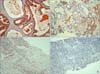
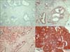
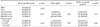
Positive staining in the cytoplasm of tumor cells was defined as reddish-brown staining in at least 10% of the tumor cells in a section. A semiquantitative scale of 0 to 3 was used to score the reactivity the samples. The immunoreactive score was determined by the percentage of positive cells and the staining intensity, ranging from no detectable signal (0) to a strong signal seen at lower-power magnification (3). Each image was scored as 0 (0%, negative), 1 (10-30%, weak), 2 (30-70%, moderate), or 3 (70-100%, strong) and classified as low expression (score 0 and 1) and high expression (score 2 and 3) (Fig. 1, 2).
4. Statistical analyses
We examined the difference in S100A2 and S100A4 expression between BPH and CaP samples among the groups, which were classified based on localization of CaP. Biochemical progression-free survival was determined based on levels of prostate-specific antigen (PSA) and studied by use of the Kaplan-Meier product limit analysis. Differences between the groups were examined by use of the log-rank test. Biochemical recurrence was defined as an increase of 0.2 ng/ml. Comparisons of means within groups were made using the one-way ANOVA test. Comparisons of immunostaining scores within groups were made using the Kruskall-Wallis test. Sample correlations were estimated using Spearman's rank correlation. Statistical significance was defined as <0.05. All analyses were performed using the Statistical Package for Social Sciences (SPSS), version 12.0 for Windows.
RESULTS
1. Comparison of S100A2 and S100A4 expression in BPH and CaP
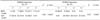
The age of the patient did not differ significantly between patients with BPH (68.28±8.05 years) and CaP (68.69±8.26 years) (p=0.082) (Table 1). We examined the expression of S100A2 and S100A4 in benign and cancer tissues. We observed that the expression of S100A4 was significantly higher than that of S100A2 in CaP tissues (p<0.05) (Table 2). In BPH tissues, the expression of S100A2 was higher than that of S100A4 (p<0.05) (Table 2).
2. Association between S100A2 and S100A4 expression and clinical stage and pathologic grade (Gleason score)
The age and follow up periods of patients did not differ significantly among the groups of CaP patients (p>0.05). We observed that the PSA level of patients was significantly different among groups according to clinical stage (p=0.023), but not significantly different among groups according to pathologic grade (p=0.264) (Table 1).
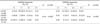
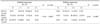
A significant progressive increase in S100A4 expression and decrease in S100A2 expression was observed in cancer specimens according to clinical stage and the pathologic grade (Gleason score) (p<0.05) (Table 3, 4, Fig. 1, 2). The Spearman correlation between S100A2 and S100A4 was found to be - 0.55 (p<0.001) based on mean that the total of every specimen.
3. Association of S100A2 and S100A4 with biochemical relapse-free survival
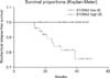
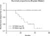
Of the 67 patients with CaP, 10 (14.9%) had evidence of biochemical relapse and 4 (5.9%) died from prostate cancer. The mean follow-up period in our study was 32.3 months. The length of biochemical relapse-free survival among the 18 patients with immunoreactive scores of 2 or 3 (Group 2) for S100A2 was statistically longer than among the 49 patients with immunoreactive scores of 0 or 1 (Group 1) (log-rank, p=0.035) (Fig. 3). The length of biochemical relapse-free survival among the 26 patients with an immunoreactive score of 0 or 1 (Group 3) for S100A4 was statistically longer than among the 41 patients with an immunoreactive score of 2 or 3 (Group 4) for S100A4 (log-rank, p=0.021) (Fig. 4).
DISCUSSION
The S100 proteins are small acidic proteins (10-12 kDa) that are found exclusively in vertebrates [19]. With at least 25 members of this protein family identified to date in humans, the S100 proteins constitute the largest subfamily of EF-hand proteins. Of these, 21 family members (S100A1 - S100A18, trichohyalin, filaggrin and repetin) have genes clustered at chromosome locus 1q21; other S100 proteins are found at chromosome loci 4p16 (S100P), 5q14 (S100Z), 21q22 (S100B), and Xp22 (S100G). First identified by Moore in 1965, the S100 proteins exhibit 25-65% identity at the amino acid level and contain 2 EF-hand motifs flanked by conserved hydrophobic residues separated by a linker region [19,20]. The sequences of the linker region and the C-terminal extension are the most variable parts of the sequence among the S100 proteins. The S100 proteins are hypothesized to participate in signal trans duction pathways that regulate cell cycle progression and differentiation; however, the precise functions of these proteins are unknown.
The S100A2 gene encodes a protein of 99 amino acids. The gene is located on chromosome 1q21, in a region that is frequently rearranged in a number of human cancers [5,8]. Expression of S100A2 is regulated during the cell cycle, with its levels increasing as cells enter the S phase, and induced by growth factors in the early G1 phase of the normal cell cycle [8]. Because S100A2 is believed to be regulated by the tumor-suppressor p53, induction of p53 activity by cell cycle arrest caused by DNA damage results in increased S100A2 transcription [21,22]. The loss of S100A2 expression is associated with the development and progression of some human cancers [9-14]. Recently, S100A2 was proposed to be a class II tumor suppressor gene because of its loss of expression in a large number of tumors. This loss of expression is believed to influence the regulation of genes that are important for normal cell growth and differentiation [12,23]. In our study, we observed decreased S100A2 expression in CaP. It is important to emphasize that we observed decreased S100A2 expression only in CaP and not in BPH (nonmalignant).
Another member of the S100 family of proteins is S100A4, which influences cell cycle progression and cell motility, and modulates intracellular adhesion and invasiveness [15,16]. Expression of the S100A4 gene has been linked to invasion and metastasis of cancer cells and is upregulated in a number of human cancers [16,24]. Amplification or overexpression of S100A4 in patients with breast cancer reflects increased metastatic potential of the cancer, which is prognostically significant and closely correlated with death [25]. S100A4 has been reported to be expressed in 44% of foci of carcinoma within colon adenoma specimens and 94% of those of colon carcinoma specimens [26]. In our study, we observed high expression of S100A4 in invasive CaP. We also observed that decreased expression of S100A4 gene reduces the growth and proliferative potential of CaP. These results indicate that the S100A4 protein may be associated with proliferation, invasion, and metastasis during the progression of CaP.
During about 5 years of follow-up, 10 of 67 (14.9%) patients had a biochemical relapse based on PSA levels. We found that the length of biochemical relapse-free survival was longer in those patients with higher expression of S100A2 and shorter for those with higher expression of S100A4. This finding indicates that expression of S100A2 and S100A4 is significantly associated with prognosis.
Our study was limited by its retrospective design, the relatively small number of patients, and the short length of follow-up. This makes it difficult to draw definitive conclusions. Because a single pathologist assessed the immunohistochemical staining of samples in a blinded fashion, interobserver variance was not tested in this study.
CONCLUSIONS
We found an inverse relationship between expression of S100A2 and S100A4 protein in CaP and BPH. Reduced expression of S100A2 and increased expression of S100A4 in CaP was associated with clinical advancement and biologic aggressiveness of tumors. Hence, the simultaneous analysis of S100A2 and S100A4 expression in prostate tissues may be a useful prognostic marker for CaP.
 XML Download