

 PDF
PDF ePub
ePub Citation
 Print
Print


INTRODUCTION
There are controversies surrounding the role of diuretic renal scans when deciding on conservative therapy or surgery in children with ureteropelvic junction obstruction (UPJO). It remains difficult to choose an optimal time for surgery as a result of the high variability in renal function, the degree of obstruction, the extent of damage, and the potential of regeneration in a growing kidney [1,2]. In addition, the relative paucity of collagen in the neonatal renal pelvis helps to alleviate the effect of high obstructing pressure [1]. However, an unrecognized obstruction may result in renal damage and renal failure. To date, diuretic renal scans provide a reliable diagnostic tool for guiding patient management. However, the value of this investigation in children has been questioned, because of its inherently high false-positive and false-negative rates [3]. Specifically, false-negative results are clinically important because they can result in missed optimal surgical opportunities. More reliable assessment tools are therefore required to aid in decision making regarding the optimal surgical time.
We retrospectively compared a conventional differential renal function (DRF) measurement with a new DRF measurement that assesses the renal parenchymal areas from imaging studies in children who underwent pyeloplasty due to unilateral UPJO.
MATERIALS AND METHODS
From September 2001 to January 2008, 29 children underwent pyeloplasty due to unilateral UPJO, and 99mTc-diethylenetriaminepentaacetic acid (99mTc-DTPA) renal scans and other imaging studies, such as magnetic resonance imaging (MRI), computed tomography (CT), and renal ultrasonography, were performed.
Diuretic renal scans were performed by using the standardized 99mTc-DTPA protocol, as a result of a discussion between the Society for Fetal Urology (SFU) and the Pediatric Nuclear Medicine Council. On the morning of the study, oral fluids were encouraged, followed by intravenous administration of 15 ml/kg of a 0.9% sodium chloride solution 30 min preceding the scan. A renal scan using 99mTc-DTPA was then performed under urinary bladder catheterization. The dosage administered was scaled for body weight and was based on an adult dose of 600 MBq. Intravenous furosemide (1 mg/kg) was given when maximum pelvicaliceal distention was observed. This usually occurred between 20 and 30 min after administration of 99mTc-DTPA [2,4].
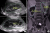
We proposed a novel method to calculate the renal parenchymal area and make correlations with DRF as measured by renal scans. All imaging studies such as MRI, CT, and renal ultrasonography were viewed on the Picture Archiving & Communications System (PACS), and all areas of measurement were conducted with the electronic drawing tool provided by the PACS radiographic software. A dedicated urologist conducted all the measurements of the renal parenchymal areas (unit areas). The unit areas of both kidneys were measured by manual tracing of the renal system excluding the extrarenal-pelvic area (region of interest) on the PACS workstation (Fig. 1). In order to obtain reliable results, we rechecked the images three times in a magnified view (×2), and an average was taken for each set of results.
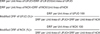
We applied the equation, DRF per unit area of UPJO (or normal contralateral kidney; NCK)=DRF of UPJO (or NCK)/ renal parenchymal area of UPJO (or NCK). Modified DRF of UPJO was calculated by using the equation, modified DRF of UPJO (or NCK)=DRF per unit area of UPJO (or NCK)×100/(DRF per unit area of UPJO+DRF per unit area of NCK) (Fig. 2).
The data were further analyzed with calculations using the McNemar chi-square test and generalized estimation equation for comparison of the modified DRF group and the conventional DRF group. All statistical tests were evaluated at a 0.05 significance level. The statistical analyses were performed by using SPSS (version 12.0; SPSS Inc, Chicago, IL, USA) computer software.
RESULTS
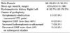
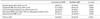
We reviewed the diuretic renal scan results of 29 pediatric patients (26 males and 3 females) who underwent pyeloplasty due to unilateral UPJO (23 left kidneys and 6 right kidneys). The mean patient age was 42.6±52.6 months (range, 3-198 months). Indications for pyeloplasty were recurrent urinary tract infection or flank pain (11 children), DRF of less than 40% on the affected side with severe hydronephrosis (8 children), progressive dilatation on a serial ultrasound (7 children), and a greater than 10% decrease in DRF on a serial renal scan (3 children) (Table 1). The mean cross-sectional areas of the UPJO kidney and of the normal contralateral kidney were 62.1±29.2 cm2 and 41.3±22.5 cm2, respectively (p<0.01). The conventional and modified DRF of the UPJO kidney were 45.2±9.2% and 35.2±9.5%, respectively (p<0.01).
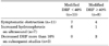
Children were divided into 2 groups on the basis of the results of the initial DRF: group I (n=8) had DRF less than 40%, and group II (n=21) had DRF greater than 40%. Thirteen children (62%) who initially belonged to group II (n=21) were reclassified into group I by the modified DRF measurement. In group II, 7 of 11 children (63.6%) whose modified DRF value was less than 40% had recurrent urinary tract infection or flank pain. A total of 6 of 7 children (86%) showed progressive dilatation on the serial ultrasound (Table 2). Table 3 compares the conventional DRF and modified DRF of the affected kidneys. The modified DRF measurement demonstrated higher accuracy than the conventional method in DRF assessment, with respect to signs and symptoms, reduction in renal function, and hydronephrosis. Modified DRF was statistically significantly different from conventional DRF (p<0.05). The false-negative rates of conventional DRF and modified DRF were 72.4% and 27.6%, respectively.
DISCUSSION
In the pediatric population, congenital urinary tract obstruction is the most common fetal anomaly identified in prenatal screening of pregnant women. It is one of the major causes of renal damage in young children [2,5]. Koff proposed that ureteral obstruction be defined as a functional or anatomical obstruction of urine flow from the renal pelvis to the ureter that results in renal damage or manifests as clinical symptoms such as recurrent urinary tract infection and flank pain when left untreated [5]. It is well known that the glomerular filtration rate (GFR) is lower in newborns than in older children, and the GFR increases several times during the initial 6 months of life. In this period, untreated obstruction can lead to early renal atrophy and permanent loss of renal function [6-8]. In addition, renal immaturity may lead to misinterpretations during preoperative and postoperative evaluations.
Diuretic renal scans have become a popular method for differentiating between obstructive and nonobstructive hydronephrosis [3,9,10]. However, the value of this investigation in children has been questioned as a result of the inaccurate results it entails [3]. To obtain maximum benefits from diuretic renal scans, intravenous hydration should be combined with diuretic administration in order to maximize urine output. Factors such as adequate hydration and diuretic use are crucial in overcoming the reservoir or 'mixing chamber' effect, which may stimulate obstruction in dilated but otherwise unobstructed systems. Consequently, standardized investigation protocols are required with the diuretic renal scan [2,4]. Adequate hydration must be ensured, and there must be sufficient residual renal function to enable diuretic response in order to define the distensibility and volume of the collecting system. Urinary bladder volume and drainage can also affect the response pattern and the clinician's ability to interpret lower ureteric drainage, which explains the use of bladder catheter drainage during the study [2,4]. Nam and Lee emphasized that the factors that help to determine true obstruction, such as renogram curves, diuretic half-lives, serial renal imaging scans, and DRFs, should be taken into account when determining the optimal surgical time in children with UPJO [11].
Another problem with DRF is the so-called supranormal renal function. It remains unclear whether this supranormal function of the obstructed kidney reflects a true increase or merely a measurement error [12,13]. The relatively high incidence (9% to 21%) of this paradoxical function is clinically important because management of hydronephrosis with supranormal function has not been clearly established to date. In our study, supranormal function (55% or greater) was present in 4 patients (13.8%). Ham et al hypothesized that supranormal DRF may occur as a result of increased renal blood flow caused by altered renal hemodynamics [14]. Consequently, there are pressing clinical needs for a more reliable test to assess the appropriateness of surgical intervention in children with UPJO.
To our knowledge, the correlation between differential parenchymal areas on imaging studies and DRF reported on renal scans has not been reported previously. Feder et al suggested that renal parenchymal areas measured by CT strongly correlate with the results of the renal scans [15]. The overall averaged difference in calculating differential function by CT versus that of renal scan was only 4.73% [15]. According to these results, measurement of DRF in kidneys with a significant size difference could be riddled with pitfalls. We propose a new methodology: DRF on the renal scan is proportional to the renal parenchymal area on imaging studies, and DRF per unit area is more accurate. In addition, kidney dimensions can be easily measured on imaging studies and the treating clinician can rapidly assess the degree and site of obstruction. Modified DRF was significantly different from conventional DRF. We reviewed 29 children with UPJO who underwent pyeloplasty, and as intraoperative findings demonstrate the most reliable diagnostic results, we suggest that there are no methodological problems comparing the false-negative results of conventional DRF with that of modified DRF: the false-negative rates of conventional DRF and of modified DRF were 72.4% and 27.6%, respectively. Furthermore, 86% of children with progressive dilatation on the serial ultrasound demonstrated DRF of less than 40% on modified DRF in group II. These results indicate that modified DRF may be a significant predictor of surgical intervention. Modified DRF measurement according to cross-sectional area showed higher diagnostic accuracy, and it may be considered a valuable method for deciding on pyeloplasty in equivocal circumstances.
There is still much debate over how best to manage obstructions in neonates. Early in the debate, a number of authors advocated early intervention to preserve renal function. There is a risk of deteriorating renal function in the future despite eventual spontaneous improvement or resolution of hydronephrosis. In addition, there is a possibility of refining our diagnostic armamentarium to detect renal decompensation at a reversible stage before the kidney becomes permanently damaged [16]. However, until now, evidence that suggests surgery will improve renal function or at least prevent further renal damage is lacking [17,18]. Increasingly, observation has been recommended for most infants, as many appear to do well without aggressive surgical intervention, and the current trend in the treatment of patients with unilateral UPJO is nonoperative care [17,18]. Koff and Campbell initially observed and subsequently performed surgery in patients with renal function and DRF deterioration [19]. They reported a study in which 104 neonates with unilateral UPJO were managed conservatively and followed up for over 5 years [20]. Only 7% of children required pyeloplasty due to DRF deterioration [20]. However, relief of obstruction is more suitable in the following conditions: DRF of less than 40% or functional reduction at follow-up, recurrent urinary tract infection despite prophylactic antibiotics treatment, or a strong likelihood of recurrent urinary tract infection regardless of the DRF value. Surgery may help to prevent renal parenchymal infection and irreversible renal damage [21-24]. Moreover, the procedure should not be delayed when indicated, because the surgical risks of pyeloplasty in infants are not as high as those of ureteral re-implantation. Our indications for pyeloplasty were recurrent urinary tract infection or flank pain (11 children), DRF of less than 40% on the affected side (8 children), progressive dilatation of hydronephrosis (7 children), and a greater than 10% decrease in DRF on serial renal scans (3 children).
This study was limited by the fact that it was performed retrospectively, and the data were analyzed in selected children who underwent pyeloplasty due to unilateral UPJO. As a consequence, we were not able to analyze false-positive results and specificity. Furthermore, measurements of the unit area were not made with a single imaging tool and therefore measurement error was possible. Finally, our study had a small sample size of 29 children; therefore, additional confirmatory studies are required in the near future.
CONCLUSIONS
Currently, DRF is one of the most important parameters applied to determine the optimal time for surgical intervention for UPJO in children. However, the value of this investigation in children has been questioned because of its high false-positive and false-negative rates. We suggest a modified DRF measurement that takes into account cross-sectional areas. Our modified DRF measurement exhibited a lower false-negative rate and may become a valuable method for deciding on pyeloplasty in children with UPJO in equivocal circumstances.
 XML Download