

 PDF
PDF ePub
ePub Citation
 Print
Print


INTRODUCTION
Cryptorchidism (from the Greek kryptos, meaning "hidden," and orchis, meaning "testis") refers to the absence of a testis from the scrotum. Isolated cryptorchidism is the most common congenital anomaly of the male genitalia, affecting almost 1% of full-term infants at the age of 1 year [1]. During embryonic life, the testes form beside the mesonephric kidneys and descend via the inguinal canal to the scrotum. If this process is faulty, a cryptorchid testis may halt along the normal path of descent (undescended or retractile testis), may travel off the normal path of decent (ectopic testis), or may die or never develop (absent testis).
The history of the study of cryptorchidism and the first attempt to correct it began in the 18th century. Like other areas of medicine, which is a combined action of art and science, the progress of techniques in orchiopexy has been supported by an improved understanding of cryptorchidism. In this context, this review aimed to describe the historical landmarks in the progress of orchiopexy in parallel with the growing knowledge of cryptorchidism that had spurred the technical advances in orchiopexy.
THE ERA OF PIONEERS
The theoretical bases that justify orchiopexy in patients with cryptorchidism originated from some critical observations by two pioneers in the 18th century, Baron Albrecht von Haller and John Hunter. Over the ensuing years, theories on the mechanism of descent, and study of the histological and physiological alterations in the cryptorchid testis came out, aiding the development of orchiopexy.
Baron Albrecht von Haller, who became the Chairman in Anatomy and Surgery at Göttenger University in the 1730s, described the abdominal position of the fetal testis in his famous work 'Opuscula Pathologica,' published in 1755. In the chapter regarding congenital hernia, he accurately indicated the presence of abdominal testis, though he did not know the exact timing of testicular descent. Also, his explanation of the phenomenon responsible for the descent of the testis was wrong. However, it was important that his description attracted the interest of John Hunter, who later made some great observations that still hold true today.
John Hunter is known as one of the fathers of modern surgery and anatomy. He was born on a small farm near Glasgow, Scotland, in 1728, the youngest of 10 children. Among his 10 siblings, he and his older brother William made a significant contribution to medicine. They founded the first independent anatomical school in London. John Hunter was a brilliant anatomist with unending curiosity, a variety of interests, and a colorful character. Although William wanted his younger brother to be a classic gentleman, his character made him a house-surgeon and later a partner in the anatomy school. He was the leading expert in the study of comparative anatomy, infectious diseases, and gunshot wounds [2,3].
John Hunter began to study on the descent of fetal testis with an interest in Baron von Haller's observations. In 1762, Hunter confirmed the abdominal position of the fetal testes as well as the neurovascular supply and the cremaster or musculus testis (a more proper name, according to Hunter). Through postmortem dissection, he first observed that the testis descends generally around the eighth month. He also described the retractile testes, undescended testes, and testicular ectopia. He did not agree with the current suggestion that the testis was forced into the scrotum by the compressive force of respiration, or pulled by the cremaster muscle. Instead, he proposed the significance of gubernaculum as a helm or rudder for testicular descent [4].
Although the precise cause of failure to descend has not been clearly understood at this time, his association of cryptorchidism with faulty testis appears interesting, as he stated that: "when one or both testicles remain through life in the belly, I believe that they are exceedingly imperfect and probably incapable of performing their natural functions, and that this imperfection prevents the disposition for descent from taking place."
He believed in the necessity of treatment of undescended testis after a period of patient observation. He wrote "As this progress is very slow, especially when the testicle is creeping through the ring, a doubt often arises whether it is better entirely to prevent its passage or to assist it by exercise or other means; and it would certainly be the best practice to assist it, if that could be done effectually and safely."
Hunter's accurate description of fetal testicular descent made great progress in the understanding of cryptorchidism. Although testicular decent and maldescent are now being explored on the molecular level, most of our knowledge on cryptorchidism is still based on his observations. Furthermore, even though many theories such as abdominal pressure, endocrine factors, cremasteric muscle contraction and gravity have been proposed to explain testicular descent, none is more important than the gubernaculum as Hunter claimed more than 2 centuries ago.
After the great discovery of Hunter, the discussion of the nature of the gubernaculum, the role of the cremaster, and the process of descent continued.
In 1866 Thomas B. Curling summarized what was known at that time regarding undescended testicles in his book, A Practical Treatise on the Diseases of the Testis [5]. Some of his observations hold true today, such as abnormal testicular function in undescended testis and proper evolutionary time limits to recovery of retained testicles. He also summarized the possible cause of the retained testicle: either the defective development of the cremasteric muscle, adhesions secondary to peritonitis, or a contracted external ring. Although he reported some misconceptions in current understandings, his observations surely gave a theoretical basis on which later doctors attempted the first orchiopexy.
UNDERSTANDING AND CONTROVERSIES ABOUT GUBERNACULUM
The structure of the gubernaculum, or genitoinguinal ligament, was first described and named by Hunter in 1762. Although most authors believe that it contributes significantly to testicular descent, there is little consensus on the mechanism involved. Controversies around gubernaculum were related to its tail structures and its role in the development of ectopic testis.
In 1840, Curling noted the gubernaculum to be a soft, solid protruding body that varied in shape and size at different stages of testicular descent [6]. He also claimed that the gubernaculum terminated in three muscular processes, which he could trace before, during and after testicular descent. He listed these as (i) external - connected to Poupart's ligament; (ii) middle - through the external ring to the bottom of the scrotum; and (iii) internal - to the os pubis and the rectus muscle sheath.
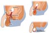
Since the gubernaculum contains muscle that is likely to contract, this led Lockwood to propose the 'traction theory' of testicular descent. Following the examination of eight fetuses (7 weeks' gestation to full term), he noted the change of distal gubernaculum from a soft, jelly-like mass into a leash of fibers that spread out to several areas and pulled the testis into the scrotum [7]. Sometime later, his description of the fibers became known as the 'tails of Lockwood' and the presumed cause of testicular ectopia (Fig. 1) [8]. Although Lockwood himself did not mention that abnormal development of the gubernacular tail could be the cause of testicular ectopia, some authors referred to Lockwood's work as an explanation for testicular ectopia [9-11].
In the early 20th century, the concept of gubernacular tails as a cause of testicular maldescent prevailed. Coley found many similarities between Curling's and Lockwood's theories and tried to amalgamate both theories [12].
Some authors reported findings in conflict to the "tails of Lockwood." Sonneland could not find any corroborative evidence either embryological or anatomical for the existence of multiple processes [9]. Instead of multiple gubernacular tails, he claimed a singular gubernacular process and believed that the fibrous attachment of ectopic testis was just a result and not the cause of testicular maldescent. McGregor also failed to find any subdivision of the gubernaculum and concluded that Lockwood's theory was unproved and proposed the 'third inguinal ring' or congenital fascial packets or barriers as a cause of testicular ectopia [13,14].
Backhouse proposed his own theory regarding the gubernaculums [15]. He described the gubernaculum as consisting only of mesenchyme (not muscles and fibers, as previous believed), which formed a column in the abdomen at taching the testis to the inguinal region and then to the floor of the scrotum. His theory was that the gubernacular mesenchyme could be disrupted by surrounding fibrous tissues to cause undescended or maldescended testis. He proposed that 'a fibrous band is formed (a "tail of Lockwood") which anchors the gubernaculum and later the testicular apparatus to the surrounding tissues, and, by virtue of the processus vaginalis having being formed normally elsewhere, the descending testis is diverted from its course in the direction of the band. This then is the aetiology of an ectopic testis and also the formation of the tails of Lockwood, which are a pathological feature rather than a normal component of the gubernaculum.' According to his theory, the tail of Lockwood indicated either a gubernacular process or an abnormal fibrous attachment, causing great confusion [15,16].
By the mid 1980s most investigators completely rejected the theory of multiple gubernacular tails providing traction to cause testicular descent. In 1987, Heyns published a landmark paper describing testicular descent in 178 human fetuses, the largest number of fetuses examined at the time. What he found was no inclusion of muscle in the gubernaculum and singular rather than multiple processes of distal gubernaculum [17].
At present, many studies have now shown conclusively that multiple gubernacular processes (or tails) do not exist and that the distal gubernaculum is unattached to surrounding tissues during inguinoscrotal migration in both rodents and humans. Attachment to the scrotum (or elsewhere) occurs only after normal (or abnormal) testicular descent is complete. Although some recent papers still referred to tails of Lockwood as a cause of ectopic testis and even transverse testicular ectopia, it is certainly time to dispel the myth of the tails of Lockwood as a cause for ectopic testis.
THE FIRST ATTEMPT AT SURGICAL CORRECTION OF CRYPTORCHIDISM
Before the periods when orchiopexy was widely accepted, an inguinal ascended testis was managed primarily with the use of truss or castration.
It was said the surgery for correction of undescended testis was attempted by several German doctors such as J.F. Rosenmerkel of Munich in 1820 and M.J. von Chelius in 1837 [4]. However, the first recorded attempt was performed by James Adams in the London Hospital in 1871 on an outpatient. His reason for correction was shown in the Lancet published at that time [18]. He proposed 3 reasons to operate on an undescended testis: poor scrotal development due to cryptorchidism, the risk of atrophy of the abdominal testis, and the likelihood of injury or pain associated with the abnormal location. He reported on one 11-week-old patient who was referred for an empty left scrotum, normal right testicle and an "oval swelling in the perineum, to the left of the middle line.. in front of the anus." Adams performed the orchiopexy with Curling through a 1.5 inch incision over the external ring. The spermatic cord and testicle were freed from attachments. The tunica vaginalis was uninjured and a catgut suture was used to affix the testicle into the scrotal pouch, after which the wound was closed. Owing to the high prevalence of erysipelas in the hospital, the operation was performed on an outpatient basis and the child was sent home. Nevertheless, the patient developed a wound infection on postoperative day 3 that progressed to fatal erysipelas. Adams personally performed the autopsy and concluded that "death was caused by peritonitis commencing in the tunica vaginalis and extending upwards." The importance of identifying and ligating a patent processus vaginalis during orchiopexy was not recognized at that time [18].
In the 1870s even minor surgical procedures carried a high risk of morbidity from infection. Adams concluded that "no operation should be undertaken in early life" because of the high likelihood of wound infection and subsequent peritonitis, unless the patient is "destined to become an equestrian." Because most orchiopexies at that time were conducted for a perineal ectopic testis, which could have caused significant discomfort to the horseman, he seemed to suggest orchiopexy limited to this kind of occupation.
ANNANDALE'S SUCCESS FOR 1st ORCHIOPEXY
Thomas "Tommy" Annandale was born at Newcastle-on-Tyne on February 2, 1838. At the age of 15 years Tommy began work as an apprentice to his father, and at age 18 he matriculated at the University of Edinburgh, receiving a doctorate in medicine 4 years later in 1860. For his surgical training, He remained in Edinburgh under the tutelage of James Syme. Then he served as private assistant for 10 years until Syme's death in 1870. At that time, he became acquainted with Joseph Lister, who had already been the senior assistant under Syme. Besides, he also got the idea of antiseptics by using a carbolic acid wound dressing from Lister. The concept of an antiseptic wound dressing technique, first reported by Lister, was revolutionary at that time, significantly reducing the risk of wound infection. In 1877, he succeeded Joseph Lister as the regius professor (chair) of clinical surgery at the University of Edinburgh. He occupied the surgical chair at Edinburgh for 30 years, from 1877 to 1907, maintaining an active surgical practice in general surgery, orthopedics, otolaryngology, urology, and other subspecialties [19].
On June 1877, Annandale was referred a 3-year-old boy with pain in the perineum on walking and running. Annandale described the care of this patient in 'The British Medical Journal' in 1879, detailing the first recorded successful orchiopexy [20]. The patient had a right ectopic testis palpated on ipsilateral perineum. On July 5, 1877, he did orchiopexy after freeing the testicle and gubernaculum from its attachment. Annandale credited Curling, who had attempted to perform orchiopexy with Adams, with the idea of anchoring the testis to the bottom of the scrotum, and his patient further benefited from the application of Lister's antiseptic technique as a dressing of carbolic acid (phenyl alcohol) was applied to the wound. Unlike the cases of Adams, the postoperative course was "satisfactory in every way," possibly due to the proper use of antiseptic techniques as Annandale has written "the whole of the operation was performed antiseptically."
Unquestionably, in the right place and at the right time, Annandale integrated the art and science of medicine. He recognized the importance of Lister's work and Curling's previous experiences, and fused their ideas into the first successful orchiopexy.
ESTABLISHMENT OF MODERN STANDARD ORCHIOPEXY: INTEGRATING MORE KNOWLEDGE INTO PRACTICE
Following the successful orchiopexy by Annandale, several authors refined the techniques, which helped to give birth to the current technique.
Max Schüller, in the 1881 Annals of Anatomy and Surgery, wrote an extensive treatise on undescended testis that included a description of the malignant potential of cryptorchidism [21]. In describing the surgical technique, he first advocated the division of the processus vaginalis to mobilize the spermatic cord in correction of the malpositioned testis. Additionally, he stressed the full division of cremaster upon the testicle and obstruction of the inguinal canal to reduce the re-ascent of testis. In contrast to Schüller's technique, in 1893, Leonard Bidwell, assistant surgeon of the West London Hospital, described a technique for inverting the testis to gain approximately an inch and a half of length and anchoring the testis to an external wire cage to provide continuous traction [22].
Arthur Dean Bevan was Professor and Head of the Department of Surgery at Rush Medical College in Chicago and later President of the American Medical Association and the American Surgical Association. In 1899, He brought the Schüller's concepts, such as division of the processus vaginalis, to the United States for the first time and took another step further, emphasizing the tension-free mobilization of the testis to the scrotum by releasing the spermatic vessels to the retroperitoneum and possible resection of them to gain further length [23]. He claimed that the need for spermatic vessel division would be lessened after increased study and experience. He also described sewing the deep layer of the superficial fascia to the aponeurosis of the external oblique to prevent retraction of the testis, using a purse-string technique. With these kinds of modifications, he reported his results in over 400 cases with an overall success rate of approximately 95% [24].
Bevan also recommended the early correction of undescended testis. His statement was further supported by Eisendrath, who showed a 90% occurrence of seminiferous tubule atrophy by 2 years of cryptorchidism [25]. This was followed by the landmark animal study by Carl Moore, who observed loss and recovery of testicular function after surgical cryptorchidism and orchiopexy, respectively [26].
From Bevan's work, three main issues concerning the surgical treatment of the undescended testis became apparent: the requirement for mobilization of the cord, the questionable necessity of division of the spermatic vessels to gain additional cord length, and the debate between traction and tension-free repositioning of the testis within the scrotum.
In the beginning of the 20th century, several concepts concerning cryptorchidism that are currently useful for understanding the disease were accepted, such as the prevalence of hernias, the possibility of torsion, the malignant potential of the "arrested" testis, and functional limitations of cryptorchid testis, both in terms of spermatogenesis and hormone production. These were well summarized by Eccles in a lecture named "The Anatomy, Physiology, and Pathology of the Imperfectly Descended Testis" [27,28].
Some surgeons tried to overcome the problem of a short length of spermatic cord. They fixed the testis to another site, such as the fascia lata or contralateral testis, for the purpose of possible lengthening of the cord. Torek in Newyork and Keetley in England independently reported the technique of fixation in the fascia lata in a similar period [29,30]. The testis was recommended to be kept in situ for 3 to 6 months and then detached carefully and repositioned in the scrotum. Torek also reported 64 cases of successful staged operation, which did not need to divide the spermatic vessels [29].
The concepts of continuous traction were revisited by Cabot and Nesbit of Michigan University again with the use of a rubber band and wire cage for approximately 12 days [31].
Although there is enthusiasm for one stage repair of orchiopexy, some difficult cases with short testicular cord will benefit by the judicious use of staged operations with traction of the testicular cord for the time being. This concept is still valid and is applied to some staged operations. Robert J. Prentiss of San Diego County Hospital added additional technical insight with his detailed depiction of the surgical anatomy of the spermatic vessels and the anatomic proof that relative lengthening of the spermatic cord could be achieved by division of the inferior epigastric vessels and medial displacement of the spermatic vessels [32].
The current method of testicular fixation within the subdartos pouch was first described by Schoemaker [33] in 1932 but was popularized by John K. Lattimer [34], at Columbia University, in 1957. He also worked out a way to implement gentle traction via an elastic band anchored in the vicinity of the patient's knee for 10 days.
With the inclusion of the subdartos pouch technique, the four key steps of standard orchiopexy were established just before the 1960s. The standard orchiopexy can be applied to almost all undescended testes with the exception of high undescended testes. The success rate ranges from 89% to 92%. Therefore, attention has turned to the treatment of high undescended testes which were not adequately treated by standard orchiopexy.
ESTABLISHMENT OF CURRENT ORCHIOPEXY
In 1979, Jones and Bagley suggested a high inguinal incision as the open surgical alternative for high canalicular or intraabdominal testes [35]. A transverse incision is made medial to the anterior superior iliac spine and carried down to the external oblique fascia just superior to the internal ring. This incision made it possible to approach the peritoneal cavity easier than through a standard inguinal incision. The point is the preservation of the spermatic vessels, high retroperitoneal mobilization of the spermatic vessels, and passage of the testis directly through the abdominal wall at the pubic tubercle (Prentiss maneuver). This procedure shares similar indications and surgical principles with laparoscopic orchiopexy and was the popular surgical approach before the advent of laparoscopic management of an intra-abdominal testis.
In case of a high undescended testis, the testicular artery and veins often limit the distal mobility of these testes. As mentioned earlier, attempts to divide the testicular artery were made well before the 20th century. However, a high atrophy rate precluded wide application. In 1959, Fowler and Stephens [36] studied the vascular anatomy of the testis and devised a means to repair a high undescended testis and preserve its blood supply via collateral circulation. Children with a long, looping vas that extends down the inguinal canal are the ideal candidates for this surgery, but less than one third of the children with intraabdominal testes were found to have this condition. Originally, Fowler and Stephens orchiopexy was known as a staged technique but it was further modified into a 2-staged operation with a better success rate (77% vs. 67%).
Prior to 1976, the non-palpable testis was only located by inguinal exploration. However Cortesi et al [37] first described the laparoscopy as a modality that could reveal the location of non-palpable testis. With increased experience, its indication was further expanded to therapeutic purposes. Bloom in 1991 described a procedure for staged pelviscopic orchiopexy [38]. The pure one-stage laparoscopic orchiopexy was first reported by Jordan and Winslow [39]. Therapeutic laparoscopy has the advantage of 1) high magnification and improved visualization 2) capability of extensive vascular dissection up to the origin of gonadal vessels, 3) minimal morbidity, and 4) the ability of creating a new internal ring medial to inferior epigastric vessels to achieve the straight vascular course to the scrotum.
Laparoscopic orchiopexy can be conducted as either one-stage orchiopexy with preservation of spermatic vessels or Fowler and Stephens orchiopexy. While current orchiopexy includes a variety of methods, all methods stem from the basic concepts of standard orchiopexy.
CONCLUSIONS
More than 200 years ago, the discussion of cryptorchidism began and the surgical techniques and philosophies have continued to evolve. The current technique of standard orchiopexy is the end result of evolved concepts. A study of the history of surgical management of the undescended testis sheds light on the rationale behind the current management.
 XML Download