

 PDF
PDF ePub
ePub Citation
 Print
Print


Bladder calculi are frequent in patients with voiding problems, foreign bodies in the bladder, urinary diversion, or recurrent urinary tract infections [1]. Various surgical interventions for the removal of bladder stones have been studied for centuries. Although open cystolithotomy is widely used for the removal of bladder calculi, current treatment trends are moving toward minimally invasive procedures, such as extracorporeal shock wave lithotripsy (ESWL), transurethral cystolithotripsy (TUCL), and percutaneous suprapubic cystolithotripsy (PCCL) [2]. However, surgical methods using the laparoscopic method have not been reported so far. We report here a case of two large bladder stones successfully removed by using a hybrid method combining the benefits of laparoscopic surgery and lithotripsy.
CASE REPORT
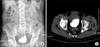
A 55-year-old man was admitted with the chief complaints of voiding difficulty, sudden stoppage of urination, frequency of urination, and residual urine sensation, which he had experienced for the previous 5 years. A pelvic radiograph showed two large radiopaque densities in the bladder (Fig. 1). Flexible cystoscopy showed two yellow and round-shaped stones at the center position, and they were not stuck to each other or to the bladder wall. The computed tomography (CT) findings showed two large stones inside the bladder, occupying more than half of its diameter (Fig. 1). One was an ovoid-shaped 5×6 cm stone, and the other was triangular in shape, 6×7 cm in size. Prostate size was 27 mg by transrectal ultrasound (TRUS). The upper tract showed no abnormality. Pyuria and Corynebacterium species were identified by urinalysis. Broad-spectrum parenteral antibiotics were given according to culture sensitivity.
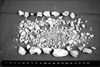
The surgery was done under general anesthesia, and the patient was placed in a supine position. An extraperitoneal approach was chosen [3]. The first (12 mm) trocar was inserted at the umbilicus by an open procedure. An extraperitoneal space was created by a handmade balloon dilator filled with 600 cc of normal saline. Then, the second (5 mm) trocar was inserted at the midline between the umbilicus and symphysis pubis. The third (5 mm) and the fourth (5 mm) trocars were inserted into the midclavicular right and left lateral abdomen at the same level as the second trocar, respectively. After removing perivesical fat, a 4 cm incision was made on the anterior surface of the bladder. Subsequently, we exposed the bladder stone by compressing the dead space of the bladder surrounding the stones with the laparoscopic stick (Fig. 2). Two large stones were put inside the laparoscopic entrapment sac that was inserted through the first trocar port site [1,4]. The stones inside the laparoscopic entrapment sac were fragmented by a lithoclast. We were able to isolate the stones from the surrounding structures as the stone was hanging from the abdominal wall; all the procedures were done with visual digital guidance. The stone's position and status were constantly checked with digital manipulation so that the sac would not be torn, and the large pieces of the stone that were sufficiently broken were immediately removed. The entrapment sac kept the fragmented pieces (which were suspected to be infected) from spreading. The bladder was sealed water-tight in two layers by Vicryl. The bladder was expanded by using normal saline to examine leakage, but no leakage was shown. It took 45 minutes to take out the stones from the bladder and to place them in the entrapment sac. It took another 90 minutes to crush and remove the trapped stones with the lithoclast. The total weight of the stones was 135 g (Fig. 3). Estimated postoperative blood loss was minimal, and no other complications were observed during or after the surgery. There was hardly any use of painkiller after surgery. The patient started diet and ambulation on postoperative day (POD) 1. Cystography on POD 14 showed no signs of urine leakage, and the urethral catheter was subsequently removed. The patient's voiding symptoms dramatically improved. Chemical analysis of the stones showed a mixed composition of magnesium ammonium phosphate (struvite).
DISCUSSION
Bladder calculi are one of the most common entities of stones occurring in the urinary tract, and, unlike kidney or ureter stones, most occur because of conditions unrelated to calculi [5]. The size and composition of the stone, the size of the enlarged prostate, the patient's conditions, a history of surgery on the lower urinary tract, the cost of the surgery, and the instruments that can be used during surgery are important factors that need to be considered in the treatment of bladder calculi [6]. It is essential that the bladder stone be removed with a minimum of trauma and damage applied to the bladder.
Although the TUCL method is more commonly used, for patients who are very young, for patients who have urethral stenosis, or when the stone burden is so large that prolonged surgery increases the risk of complications such as urethral stenosis, urethral trauma (due to taking the stone fragment out of the bladder), and visual disturbance (due to hematuria and stone dust) [7]. PCCL and sometimes open methods are preferred. In some cases, fragmented stones are difficult to remove completely by cystolithotripsy and they act as a novel nidus. In addition, when large infected stones are treated with intracorporeal cystolithotripsy, injury of the bladder wall may cause urinary tract infection and in severe cases even sepsis. Numerous, large stones such as in this case cause difficulty in stone fixation within the bladder during lithotripsy and can be a problem for the practitioner when removing other stones. When the stones are very large, 6-7 cm in size as in this case, the use of the open method will cause the window of excision to increase, which then leads to an increase in complications such as bleeding, postoperative pain, and problems in wound management [8].
We used laparoscopy with an extraperitoneal approach to prevent unnecessary injury to other organs, including the bowel. Also, we performed lithotripsy on stones trapped in an entrapment sac so that we could remove the stones completely and take them out through a small working incision. Miller and Park [4] performed modified percutaneous cystolithotomy without lithotripsy (due to a small stone size) using a laparoscopic entrapment sac under endoscopic guidance in 4 patients with augmented bladders. They mentioned that the application of this technique may minimize the risk of residual fragments. The advantage of the laparoscopic approach is that we can easily remove large stones by simply excising the bladder and exposing it as in open surgery. The benefits of using direct visual lithotripsy include being able to see the stone directly, to guide the stone with digital manipulation, to remove incomplete fragments of broken stones, and to reduce surgery time to lower the chances of bleeding. There is less of a concern for complications such as constriction of the urethra and damage to the bladder, and because the procedures are done inside the sac, there is almost no chance of infection caused by the fragmented stones. Also, one of the important reasons for selecting this technique over TUCL is that the prostate was not enlarged to resect. However, a longer operation time than open surgery and the need for skilled surgeons with accomplished skills in laparoscopy must be taken into consideration. Unfortunately, the cause of bladder stones has not been proved.
In cases of bladder stones that are large and have a large stone burden, such as is reported in this case, our novel technique is sufficiently safe and feasible and the stones can be efficiently treated. This technique should be considered as a treatment option.

 XML Download