

 PDF
PDF ePub
ePub Citation
 Print
Print


Abstract
Purpose
We investigated pre-urodynamic study parameters of bladder outlet obstruction (BOO) defined by pressure-flow study (PFS) in female patients without anatomical obstruction.
Materials and Methods
The cohort of this study consisted of 320 women who did not have anatomical BOO in whom urodynamic study was conducted for lower urinary tract symptoms (LUTS). BOO was defined when the PFS maximal flow rate (Qmax) was ≤12 ml/sec and Pdet Qmax was ≥25 cmH2O. The main outcomes were the incidence of BOO and its predictive factors in our cohort.
Results
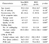
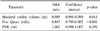
Of the total patients, 39 (12.2%) were diagnosed with BOO in the PFS. Free Qmax and maximal voided volume (MVV) were significant predictors of BOO (p<0.001, p=0.011, respectively) in the multivariate logistic regression. When free Qmax was set to ≤15 ml/sec, its sensitivity and specificity predicting BOO were 82% and 72%, respectively; when MVV was set to ≤350 ml, its values were 71% and 46%, respectively. However, the positive predictive values (PPVs) of free Qmax and MVV were low (34.4% and 28.2%, respectively), whereas the negative predictive values (NPVs) of these parameters were relatively high (96.5% and 91.2%, respectively).
Conclusions
Factors predicting BOO defined by PFS in female patients complaining LUTS without anatomical obstruction were free Qmax and MVV. The PPV of these factors was low, and the NPV was high. Therefore, if free Qmax is >15 ml/sec or MVV is >350 ml, PFS may be not essential. On the contrary, if free Qmax and MVV are below these levels, PFS may be indicated to evaluate the presence of BOO.
Figures and Tables
Fig. 1
Receiver operator characteristic curves of free Qmax and maximal voided volume, and the sensitivity and specificity for detection of BOO according to their cutoff levels. Qmax: maximal flow rate, AUC: area under curve, CI: confidence interval, BOO: bladder outlet obstruction.
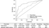
Table 1
Urodynamic study parameters in female patients with lower urinary tract symptoms according to the presence of PFS-proven BOO
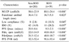
Data presented are Mean±SD or number (%). PFS: pressure-flow study, BOO: bladder outlet obstruction, MUCP: maximal urethral closure pressure, IDO: idiopathic detrusor overactivity, Qmax: maximal flow rate, Pdet, open: detrusor pressure at the onset of measured flow, PdetQmax: detrusor pressure at maximal flow rate, PVR: post-void residual, a: Student's t-test, b: chi-square test
References
1. Corrin B, Mayor D, Moor T. The pathology of bladder neck obstruction in the female patient. J Urol. 1963. 90:434–439.
2. Farrar DJ, Osborne JL, Stephenson TP, Whiteside CG, Weir J, Berry J, et al. A urodynamic view of bladder outflow obstruction in the female: factors influencing the results of treatment. Br J Urol. 1975. 47:815–822.
3. Nitti VW, Tu LM, Gitlin J. Diagnosing bladder outlet obstruction in women. J Urol. 1999. 161:1535–1540.
4. Kaufman MR, Scarpero H, Dmochowski RR. Diagnosis and management of outlet obstruction in the female. Curr Opin Urol. 2008. 18:365–369.
5. Carr LK, Webster GD. Voiding dysfunction following incontinence surgery: diagnosis and treatment with retropubic or vaginal urethrolysis. J Urol. 1997. 157:821–823.
6. Groutz A, Blaivas JG, Chaikin DC. Bladder outlet obstruction in women: definition and characteristics. Neurourol Urodyn. 2000. 19:213–220.
7. Schäfer W, Abrams P, Liao L, Mattiasson A, Pesce F, Spangberg A, et al. Good urodynamic practices: uroflowmetry, filling cystometry, and pressure-flow studies. Neurourol Urodyn. 2002. 21:261–274.
8. Defreitas GA, Zimmern PE, Lemack GE, Shariat SF. Refining diagnosis of anatomic female bladder outlet obstruction: comparison of pressure-flow study parameters in clinically obstructed women with those of normal controls. Urology. 2004. 64:675–679.
9. Griffiths DJ. Pressure-flow studies of micturition. Urol Clin North Am. 1996. 23:279–297.
10. Chassagne S, Bernier PA, Haab F, Roehrborn CG, Reisch JS, Zimmern PE. Proposed cutoff values to define bladder outlet obstruction in women. Urology. 1998. 51:408–411.
11. Lemack GE, Zimmern PE. Pressure flow analysis may aid in identifying women with outflow obstruction. J Urol. 2000. 163:1823–1828.
12. Blaivas JG, Groutz A. Bladder outlet obstruction nomogram for women with lower urinary tract symptomatology. Neurourol Urodyn. 2000. 19:553–564.
13. Akikwala TV, Fleischman N, Nitti VW. Comparison of diagnostic criteria for female bladder outlet obstruction. J Urol. 2006. 176:2093–2097.
14. Kim H, Lee U, Lee M, Choo MS. Cut-off value for bladder outlet obstruction in pressure-flow study in female: a prospective study. Korean J Urol. 2001. 42:1146–1151.
15. Raz S, Smith RB. External sphincter spasticity syndrome in female patients. J Urol. 1976. 115:443–446.
16. Oelke M, Baard J, Wijkstra H, de la Rosette JJ, Jonas U, Höfner K. Age and bladder outlet obstruction are independently associated with detrusor overactivity in patients with benign prostatic hyperplasia. Eur Urol. 2008. 54:419–426.
17. Im JG, Kim JC, Seo SI, Park YH, Hwang TK. Bladder outlet obstruction in female patients with lower urinary tract symptom. Korean J Urol. 2003. 44:1116–1120.
18. Kessler TM, Studer UE, Burkhard FC. The effect of terazosin on functional bladder outlet obstruction in women: a pilot study. J Urol. 2006. 176:1487–1492.
 XML Download