

 PDF
PDF ePub
ePub Citation
 Print
Print


Renal trauma accounts for 1-3% of all patients with traumatic injury.1 Arteriovenous fistula (AVF) is rare and occurs mostly after penetrating renal trauma, most commonly after renal biopsy.2,3 The incidence of AVF after renal stab wounds varies between 0% and 7% according to several studies.3-5
About 50% to 70% of cases of renal AVF occurring after renal biopsy improve without any treatment. But most post-traumatic AVFs require radiologic interventions and sometimes nephrectomy.1
Renal angiography is used for diagnosis and treatment. Among the many treatment methods, superselective embolization is the safest and most effective treatment modality when an AVF is confirmed.6
Rare complications of embolization do occur, including renal abscess, postembolization syndrome, impaired renal function, pulmonary embolism caused by migration of coils, allergic reaction, and hematoma on the arterial puncture site.7,8 When complications do occur, close follow-up is required for possible potentially life-threatening conditions.
Here we report a very rare case of a newly discovered pseudoaneurysm on an adjacent vessel 3 weeks after superselective embolization for renal AVF and pseudoaneurysm after a renal stab wound.
CASE REPORT
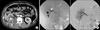
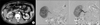
A 35-year-old man was admitted to the emergency room with gross hematuria and abdominal pain after receiving multiple stab wounds. A physical exam revealed two stab wounds on the right flank and right upper quadrant of the abdomen. The patient's mental status was alert and his blood pressure and pulse rate were normal. Abdominal computed tomography (CT) showed grade III right renal trauma, hemothorax, and suspected hemoperitoneum, but vessel injury of the right kidney was not observed. A thoracostomy was done to treat the hemothorax. Considering the patient's young age and his being hemodynamically stable, conservative management was planned for the renal trauma. Laparotomy was performed because intraperitoneal hemorrhage was suspected, but no internal organ injury was observed. However, gross hematuria persisted until 7 days after the laparotomy. The follow-up abdominal CT evaluation revealed a 1.7 cm sized pseudoaneurysm with AVF on the right lower pole of kidney (Fig. 1A). Right renal arteriography was performed by the Seldinger technique after puncturing the right pulmonary artery. As a result, we discovered AVF with pseudoaneurysm in the posterior branch of the lower pole of the renal artery. Superselective embolization with a coil (Tornado® Embolization Microcoils) was performed to treat the renal AVF with pseudoaneurysm through percutaneous renal angiography (Fig. 1B). Neither AVF nor pseudoaneurysm was observed after the procedure. No anti-coagulant was used before or after angiography. The patient was discharged without any complications following the embolization. The patient was then readmitted to the emergency room 3 weeks after the embolization for sudden gross hematuria. Abdominal CT showed no abnormality on the embolized vessel but a newly discovered pseudoaneurysm, sized 9 mm, in the interlobar artery branch of the mid-artery (Fig. 2A). Superselective embolization with a coil was performed by percutaneous renal angiography on the vessel with the new pseudoaneurysm (Fig. 2B). The patient showed no signs of complications during 6 months of follow-up after the second embolization.
DISCUSSION
Delayed hemorrhage is one of the most serious complications of renal trauma. It is common with laceration in the renal cortex, especially when conservatively managing a renal stab wound.2 The possibility of delayed hemorrhage occurring after renal-penetrating trauma or stab wound is reported to be 18% to 23%.2,5 Most of the delayed hemorrhage is thought to be caused by renal AVF.9
Direct injury on the artery due to a stab wound causes pooling of blood in the tunica adventitia, renal parenchyme, and Gerota's fascia and the development of a hematoma. When the bleeding stops, absorption of the hematoma and necrotic tissue creates a space between the tunica intima and adventitia, which forms a pseudoaneurysm. The pseudoaneurysm increases in size and may rupture into the pelvocaliceal system or perirenal space.
Most patients present with symptoms such as hematuria, flank pain, abdominal bruit on auscultation, and hypertension. The possibility of developing a pseudoaneurysm must be considered, especially when hematuria persists in patients with penetrating renal trauma or a renal stab wound.10
Angiography is the most definitive diagnostic tool for pseudoaneurysm and is now highly recommended due to the convenience of treating with embolization during the procedure.6 Complications following embolization include renal abscess, postembolization syndrome, impaired renal function, pulmonary embolism caused by migration of coils, allergic reaction, and hematoma on the arterial puncture site.7,8
The present case is very rare; there are no known reports of a newly discovered pseudoaneurysm following the embolization of renal AVF with pseudoaneurysm.
Vascular complications such as AVF and pseudoaneurysm should always be considered in renal trauma, especially in renal stab wound patients, and require angiography when associated symptoms are present. Complications may cause life-threatening conditions compared to renal injury due to blunt trauma. Therefore, continuous follow-up after the embolization is necessary to minimize the occurrence of complications. In particular, delayed bleeding or hematuria following the embolization require close follow-up for possible pseudoaneurysm, which may rarely occur, as in this case.
 XML Download