

 PDF
PDF ePub
ePub Citation
 Print
Print


Abstract
Urachal xanthogranuloma is an extremely rare disease. A 23-year-old man presented with severe lower abdominal pain and voiding frequency. Computed tomography revealed a urachal mass with bladder invasion, which was suspected to be a urachal carcinoma or abscess. Laparoscopic urachal resection was performed with a minimal incision. Histopathologic examination identified the mass as a urachal xanthogranuloma.
REFERENCES
1.Yamamoto T., Mori Y., Katoh Y., Iguchi M., Minamidari K., Sawai Y, et al. A case of urachal xanthogranuloma suspected to be a urachal tumor. Hinyokika Kiyo. 2004. 50:493–5.
2.Carrere W., Gutiérrez R., Umbert B., Solé M., Menéndez V., Carretero P. Urachal xanthogranulomatous disease. Br J Urol. 1996. 77:612–3.

3.Diaz Candamio MJ., Pombo F., Arnal F., Busto L. Xanthogranulomatous urachitis: CT findings. J Comput Assist Tomogr. 1998. 22:93–5.
4.Kuo TL., Cheng C. Xanthogranulomatous Inflammation of urachus mimicking urachal carcinoma. Urology. 2009. 73:443.
5.Tian J., Ma JH., Li CL., Xiao ZD. Urachal mass in adults: clinical analysis of 33 cases. Zhonghua Yi Xue Za Zhi. 2008. 88:820–2.
6.Navarrete S., Sánchez Ismayel A., Sánchez Salas R., Sánchez R., Navarrete Llopis S. Treatment of urachal anomalies: a minimally invasive surgery technique. JSLS. 2005. 9:422–5.
7.Wadhwa P., Kolla SB., Hemal AK. Laparoscopic en bloc partial cystectomy with bilateral pelvic lymphadenectomy for urachal adenocarcinoma. Urology. 2006. 67:837–43.
8.Kim DY., Kim HS., Kim IK., Moon I., Kim TS., Choi S, et al. Xanthogranulomatous cystitis presenting as a urachal carcinoma. Korean J Urol. 2004. 45:1180–2.
9.Han DH., Choi HJ., Kim JH., Shin JS., Chung KJ., Choi HY, et al. Xanthogranulomatous cystitis. Korean J Urol. 2004. 45:958–61.
10.Fornari A., Dambros M., Teloken C., Hartmann AA., Kolling J., Seben R. A case of xanthogranulomatous cystitis. Int Urogy-necol J Pelvic Floor Dysfunct. 2007. 18:1233–5.
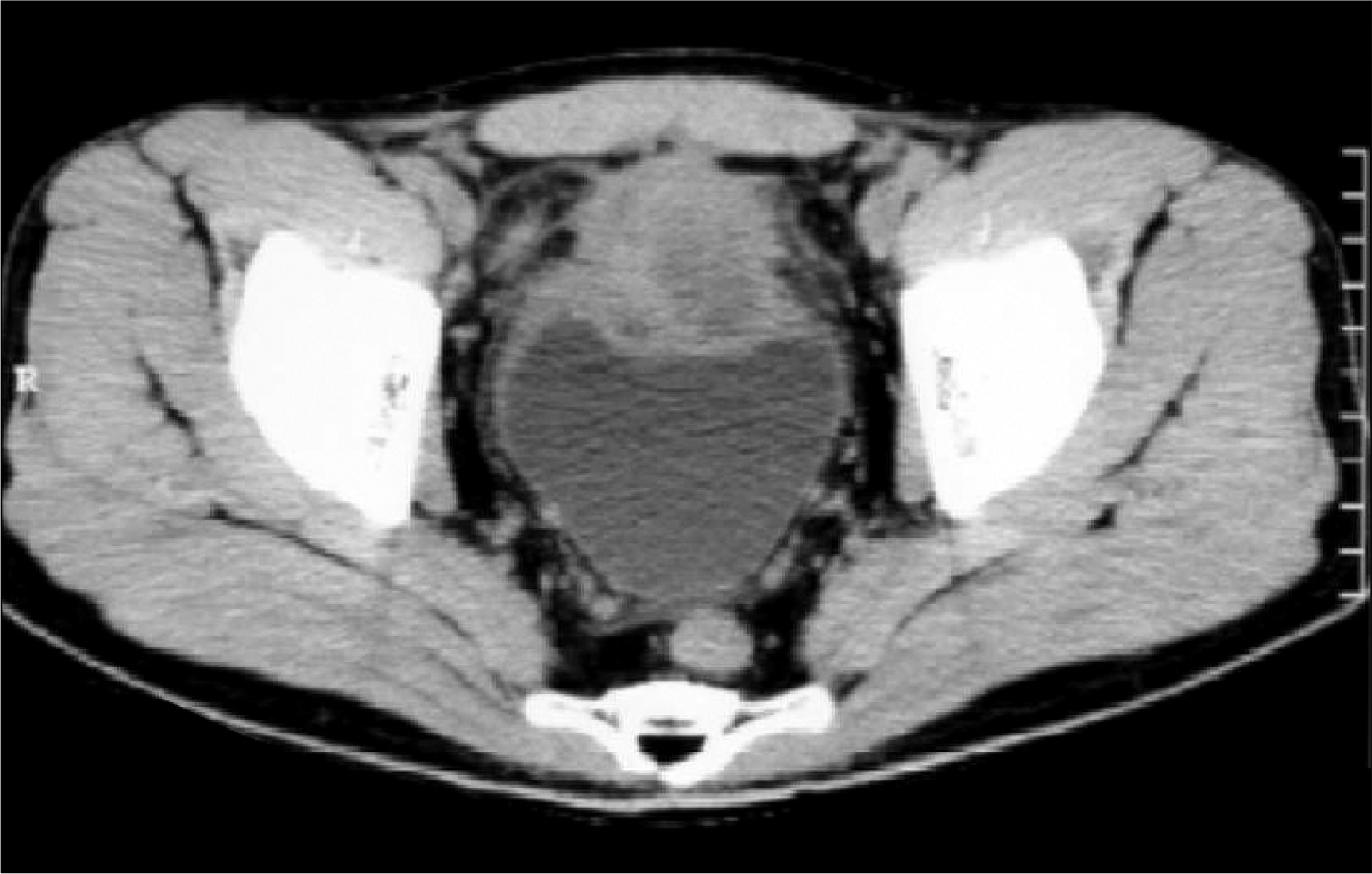
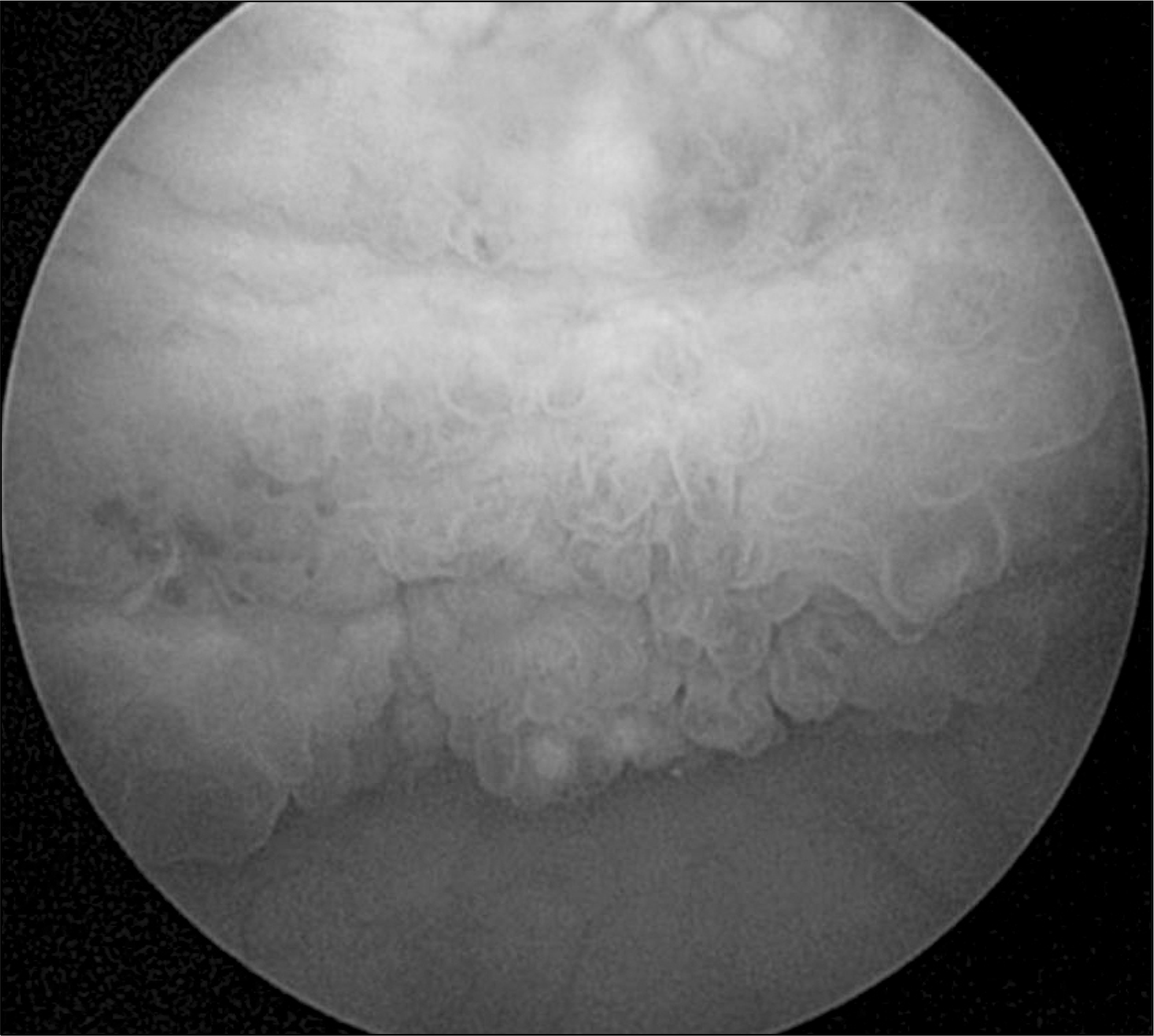
Fig. 3
Laparoscopic excision of the urachus. By use of a “twist and roll” action, the urachus was twirled around the grasping forceps.8
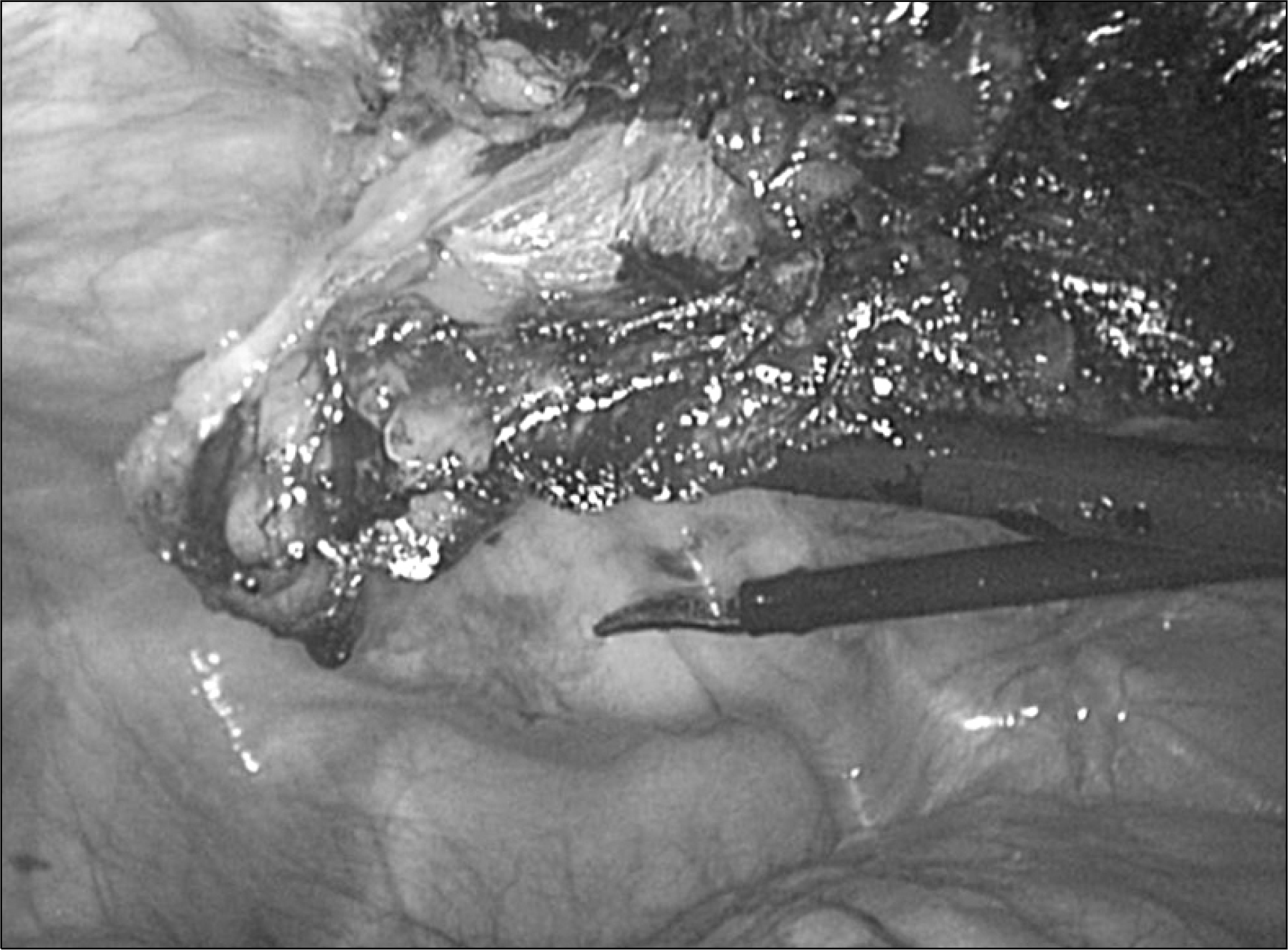
Fig. 4
(A) Gross Specimen. The mass is transected and shows a yellowish necrotic area between adipose tissues. (B) Microphotography showing urachal remnant (center) and inflammatory changes (H&E, x20). (C) Infiltration of foamy histiocytes favoring xanthogranulomatous changes (H&E, x200). The right lateral side is a smooth muscle of bladder (normal). (D) Lesion showing typical lipid-laden histiocytes with fine cytoplasmic vacuolization and scattered lymphocytes (H&E, x400).

 XML Download