Abstract
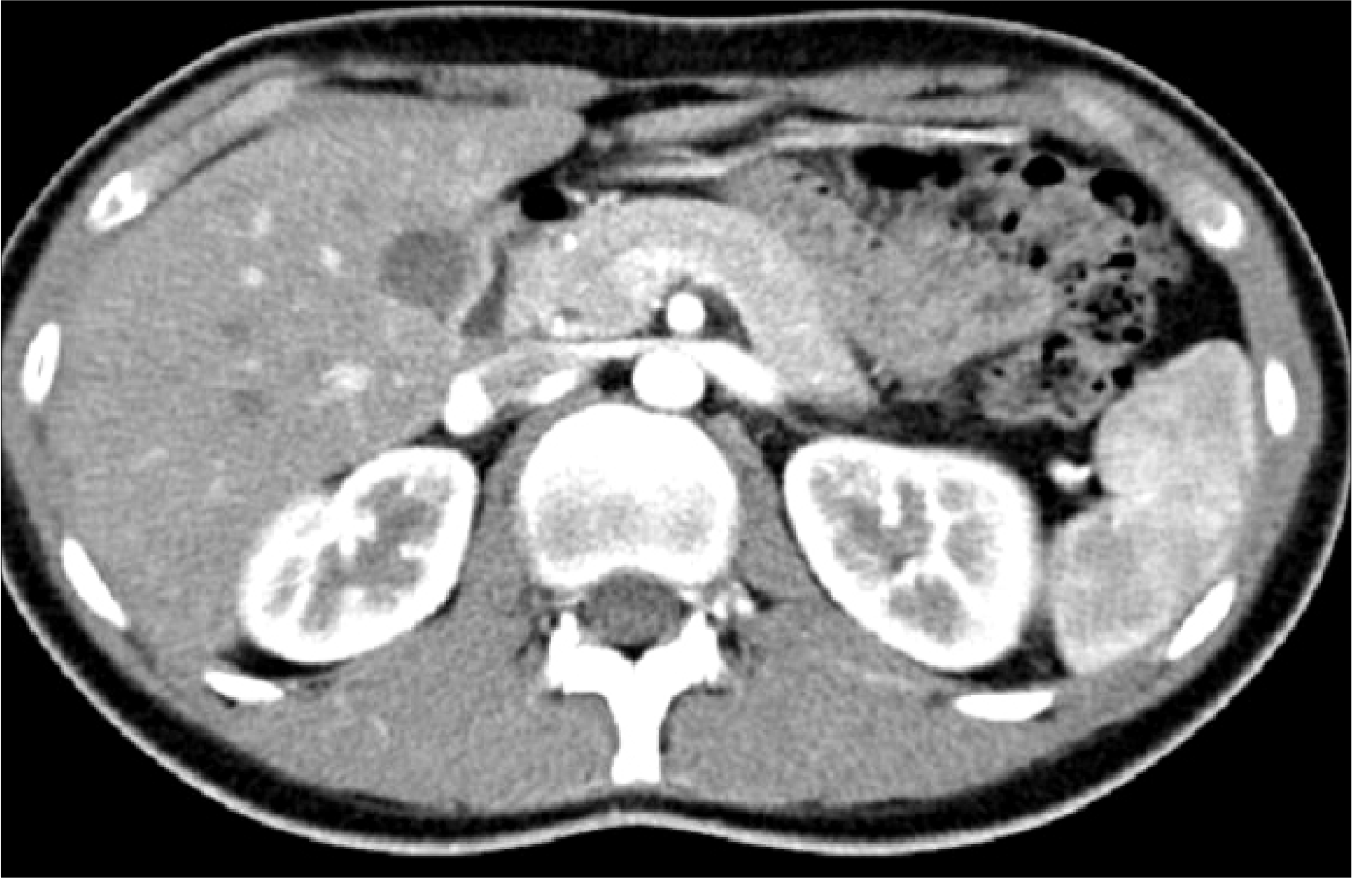
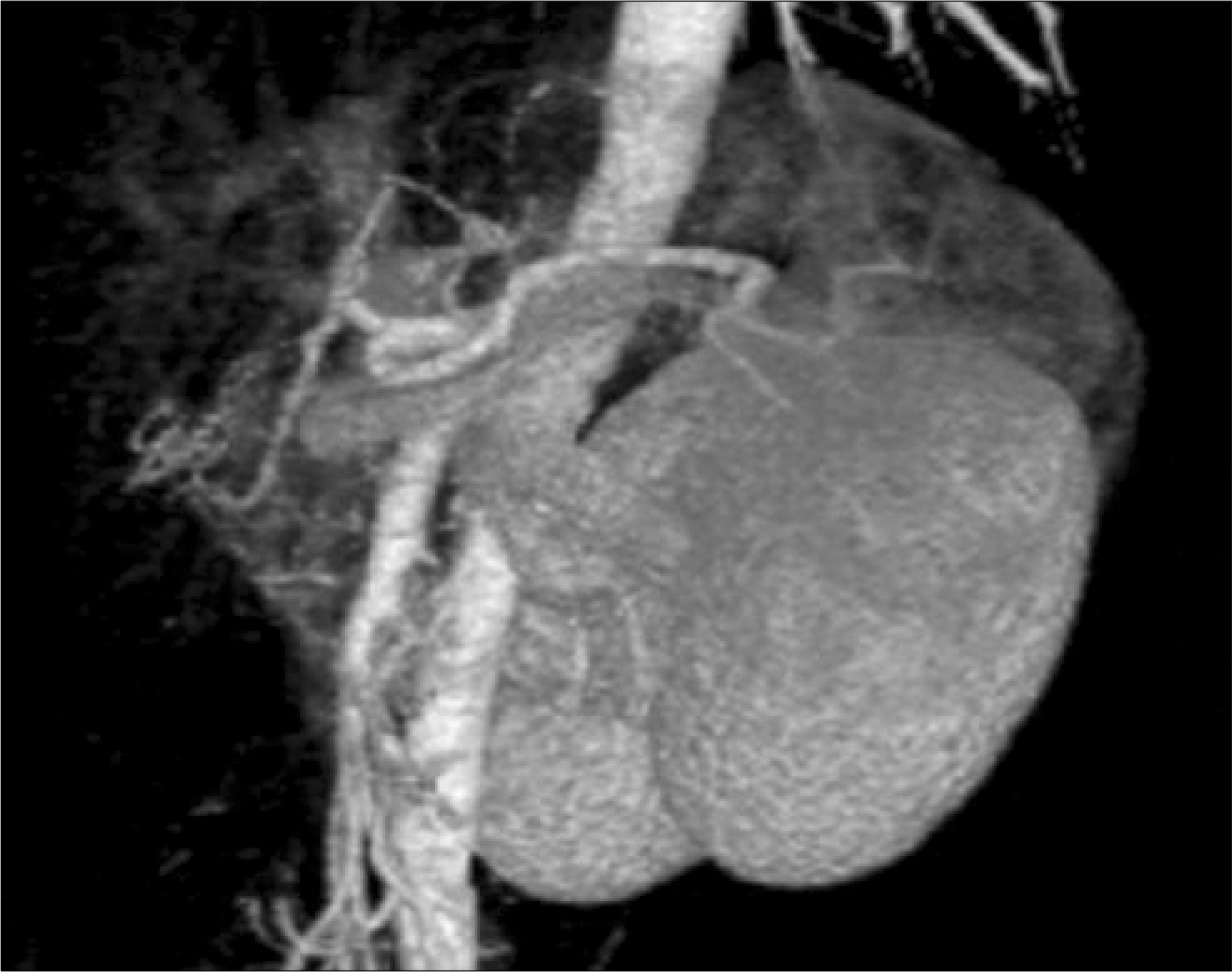
We report a case of nutcracker syndrome diagnosed with 3-dimensional computed tomography angiography (3-D CTA). Nutcracker syndrome had been confirmed by conventional venography until recent years. Nowadays, with the development of imaging techniques, color Doppler sonogram and 3-D CTA are replacing venography for the diagnosis of nutcracker syndrome. The patient, a 20-year-old male, had abrupt gross hematuria and left abdominal pain 6 months previously and intermittent microscopic hematuria thereafter. Including renal biopsy, the results of conventional hematuria study showed no abnormalities. 3-D CTA showed left renal vein compression between the abdominal aorta and superior mesenteric artery and collateral veins. The angle and distance between the superior mesenteric artery and aorta at the level of the left renal vein were 35o and 3.0 mm, respectively. We diagnosed nutcracker syndrome and later confirmed the diagnosis with venography.
Go to : 
REFERENCES
1.de Schepper A. “Nutcracker” phenomenon of the renal vein and venous pathology of the left kidney. J Belge Radiol. 1972. 55:507–11.
2.Fitoz S., Yalcinkaya F. Compression of left inferior vena cava: a form of nutcracker syndrome. J Clin Ultrasound. 2008. 36:101–4.

3.Zhang HS., Li M., Jin W., San P., Xu P., Pan S. The left renal entrapment syndrome: diagnosis and treatment. Ann Vasc Surg. 2007. 21:198–203.

4.Wendel RG., Crawford ED., Hehman KN. The “nutcracker” phenomenon: an unusual cause for renal varicosities with hematuria. J Urol. 1980. 123:761–3.

5.Rudloff U., Holmes RJ., Prem JT., Faust GR., Moldwin R., Siegel D. Mesoaortic compression of the left renal vein (nutcracker syndrome): case reports and review of the literature. Ann Vasc Surg. 2006. 20:120–9.

6.Little AF., Lavoipierre AM. Unusual clinical manifestations of the Nutcracker Syndrome. Australas Radiol. 2002. 46:197–200.

7.Fu WJ., Hong BF., Gao JP., Xiao YY., Yang Y., Cai W, et al. Nutcracker phenomenon: a new diagnostic method of multislice computed tomography angiography. Int J Urol. 2006. 13:870–3.

8.Lee SE., Cho SW., Yang SC. A case of nutcracker syndrome surgically corrected by extraperitoneal flank approach. Korean J Urol. 1996. 37:1027–30.
9.Cho SY., Shin MS., Rhee HW., Seo JM. A case of the Nutcracker syndrome. Korean J Urol. 1999. 40:120–1.
10.Yim SH., Koh JS., Kim HW., Yang CH., Jung JH., Lee JY. Expandable metallic stent placement for Nutcracker syndrome. Korean J Urol. 2004. 45:390–2.
Go to : 


 PDF
PDF ePub
ePub Citation
 Print
Print


 XML Download