Abstract
Purpose
The object of our study was to examine the clinical significance of pediatric testicular microlithiasis (TM) as it relates to testicular cancer.
Materials and Methods
Seven boys in whom TM was incidentally detected were followed for a mean of 51±44 months (range: 9-119 months) for testicular cancer surveillance. The average patient age at the initial diagnosis was 9.8±2.5 years. The frequency of coincidental TM detected on scrotal ultrasonography performed for all clinical purposes at our institution between January 1997 and January 2008 was investigated. Eighty-two testicular cancer patients and 1,006 noncancer patients underwent ultrasonography between 1997 and 2006, and these patients were divided into two age groups (children, age <15 years; adults, age ≥15 years) for purposes of analysis.
Results
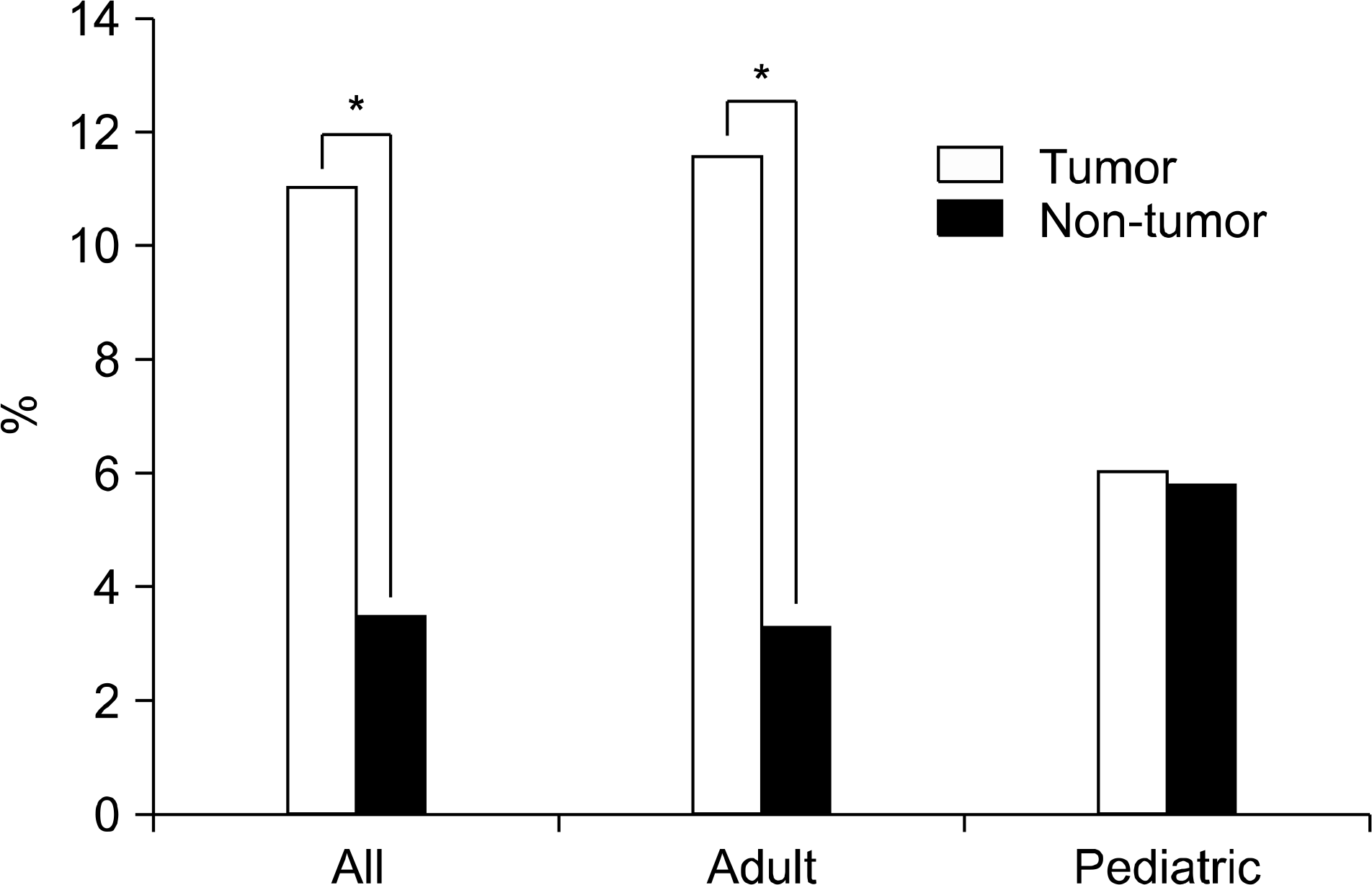
Of the seven patients followed solely for TM, none developed testicular cancer during the surveillance period. Coincidental TM seen on scrotal ultrasonography was significantly higher in the testicular cancer patients than in the noncancer controls (11% (9/82) vs. 3.5% (36/1,006), p <0.0001). According to the age groups, TM was found in 6% and 5.8% of the testicular cancer patients and the noncancer controls, respectively, in the children’s group, whereas in the adult group, 11.6% and 3.3% of the patients in the respective groups were found to have TM.
Conclusions
The incidence of testicular cancer development in children with incidentally detected TM was very low, and the incidence of coincidental TM in children with testicular cancer did not differ from that in the noncancer control patients. However, the significantly higher incidence of TM accompanying testicular cancer after puberty may suggest an association of the two pathologies, which would then mandate cancer surveillance in cases of incidentally detected TM in this age group.
REFERENCES
1.Nistal M., Paniagua R., Diez-Pardo JA. Testicular microlithiasis in 2 children with bilateral cryptorchidism. J Urol. 1979. 121:535–7.

2.Vegni-Talluri M., Bigliardi E., Vanni MG., Tota G. Testicular microliths: their origin and structure. J Urol. 1980. 124:105–7.

3.Pierik FH., Dohle GR., van Muiswinkel JM., Vreeburg JT., Weber RF. Is routine scrotal ultrasound advantageous in infertile men? J Urol. 1999. 162:1618–20.

4.Janzen DL., Mathieson JR., Marsh Jl., Cooperberg PL., del Rio P., Golding RH, et al. Testicular microlithiasis: sonographic and clinical features. AJR Am J Roentgenol. 1992. 158:1057–60.

5.Furness PD 3rd., Husmann DA., Brock JW 3rd., Steinhardt GF., Bukowski TP., Freedman AL, et al. Multi-institutional study of testicular microlithiasis in childhood: a benign or premalignant condition? J Urol. 1998. 160:1151–4.

6.Kwon JI., Kim SJ., Kim YS. Sonographically detected testiscular microlithiasis. Korean J Urol. 1997. 38:312–4.
7.Kang HW., Kang YN., Kim KS. Fifteen cases of testicular microlithiasis. Korean J Urol. 1998. 39:1259–63.
8.Fiona NM., Shantini R., Jane LC., Seahadri S., Grodon HM., Paul SS. Testicular calcification and microlithiasis: association with primary intra-testicular malignancy in 3,477 patients. Eur Radiol. 2007. 17:363–9.

9.Serter S., Gumus B., Unlu M., Tuncyurek O., Tarhan S., Ayyildiz V, et al. Prevalence of testicular microlithiasis in an asymptomatic population. Scand J Urol Nephrol. 2006. 40:212–4.

10.Ikinger U., Wurster K., Terwey B., Mohring K. Microcalcifications in testicular malignancy: diagnostic tool in occult tumor? Urology. 1982. 19:525–8.

11.Otite U., Webb JA., Oliver RT., Badenoch DF., Nargund VH. Testicular microlithiasis: Is it a benign condition with malignant potential? Eur Urol. 2001. 40:538–42.

12.Dagash H., Mackinnon EA. Testicular microlithiasis: What does it mean clinically? BJU Int. 2007. 99:157–60.

13.Ravichandran S., Smith R., Cornford PA., Fordham MV. Surveillance of testicular microlithiasis? Results of an UK based national questionnaire survey. BMC Urol. 2006. 6:8.

14.Salisz JA., Goldman KA. Testicular calcifications and neoplasia in patient treated for subfertility. Urology. 1990. 36:557–60.
15.Kocaoglu M., Bozlar U., Bulakbasi N., Saglam M., Ucoz T., Somuncu I. Testicular microlithiasis in pediatric age group: ultrasonography findings and literature review. Diagn Interv Radiol. 2005. 11:60–5.
Fig. 2.
Results according to our coincidence data. Testicular microlithiasis was found in 11% (9 of 82) of the testicular cancer patients and in 3.5% (36 of 1,006) of the non-testicular-cancer patients, thus indicating a significantly higher incidence in the testicular cancer group (p=0.0001). The incidence of testicular microlithiasis among adults was 11.6% and 3.3% in the same patient groups, but in the pediatric patients it was 6% and 5.8%, respectively. *: p<0.05.

Table 1.
Patient characteristics and reason for ultrasonography in patients with testicular microlithiasis by long-term follow-up


 PDF
PDF ePub
ePub Citation
 Print
Print


 XML Download