

 PDF
PDF ePub
ePub Citation
 Print
Print


Abstract
The rare ‘burned out’ phenomenon in germ cell tumors is known as the presence of an extragonadal germ cell tumor with a spontaneously regressed testicular tumor found in common metastatic sites, including the retroperitoneal, mediastinal, supraclavicular, cervical, and axillary lymph nodes; lung; and liver. We report a patient who presented with a retroperitoneal extragonadal germ cell tumor with a spontaneously regressed testicular tumor.
REFERENCES
1.Scholz M., Zehender M., Thalmann GN., Borner M., Thoni H., Studer UE. Extragonadal retroperitoneal germ cell tumor: evidence of origin in the testis. Ann Oncol. 2002. 13:121–4.

2.Fabre E., Jira H., Izard V., Ferlicot S., Hammoudi Y., Theodore C, et al. ‘Burned-out' primary testicular cancer. BJU Int. 2004. 94:74–8.
3.The Korean Society of Pediatric Urology. Pediatric testicular tumor registry in Korea: 1997-2001. Korean J Urol. 2004. 45:563–72.
4.Baek SH., Park JB., Jang YH., Park YW., Lee JH., Min SK. Spontaneous testicular hemorrhagic necrosis masquerading as a testis tumor. Korean J Urol. 2004. 45:962–4.
5.Hailemariam S., Engeler DS., Bannwart F., Amin MB. Primary mediastinal germ cell tumor with intratubular germ cell neoplasia of the testis-further support for germ cell origin of these tumors: a case report. Cancer. 1997. 79:1031–6.
6.Saleh FH., Crotty KA., Hersey P., Menzies SW., Rahman W. Autonomous histopathological regression of primary tumours associated with specific immune responses to cancer antigens. J Pathol. 2003. 200:383–95.
7.Tasu JP., Faye N., Eschwege P., Rocher L., Bléry M. Imaging of burned-out testis tumor: five new cases and review of the literature. J Ultrasound Med. 2003. 22:515–21.
8.Böhle A., Studer UE., Sonntag RW., Scheidegger JR. Primary or secondary extragonadal germ cell tumors? J Urol. 1986. 135:939–43.
9.Geldart TR., Simmonds PD., Mead GM. Orchidectomy after chemotherapy for patients with metastatic testicular germ cell cancer. BJU Int. 2002. 90:451–5.
10.Israel A., Bosl GJ., Golbey RB., Whitmore W Jr., Martini N. The results of chemotherapy for extragonadal germ-cell tumors in the cisplatin era: the Memorial Sloan-Kettering Cancer Center experience (1975 to 1982). J Clin Oncol. 1985. 3:1073–8.
Fig. 1
(A) Computed tomography (CT) of the retroperitoneum shows an enhanced mass (black arrow) with a lobulated margin and necrotic portion located between the pancreatic head and the inferior vena cava. (B) Proton emission tomography (PET) shows 2 hypermetabolic masses between the duodenum and the inferior vena cava (white arrow).
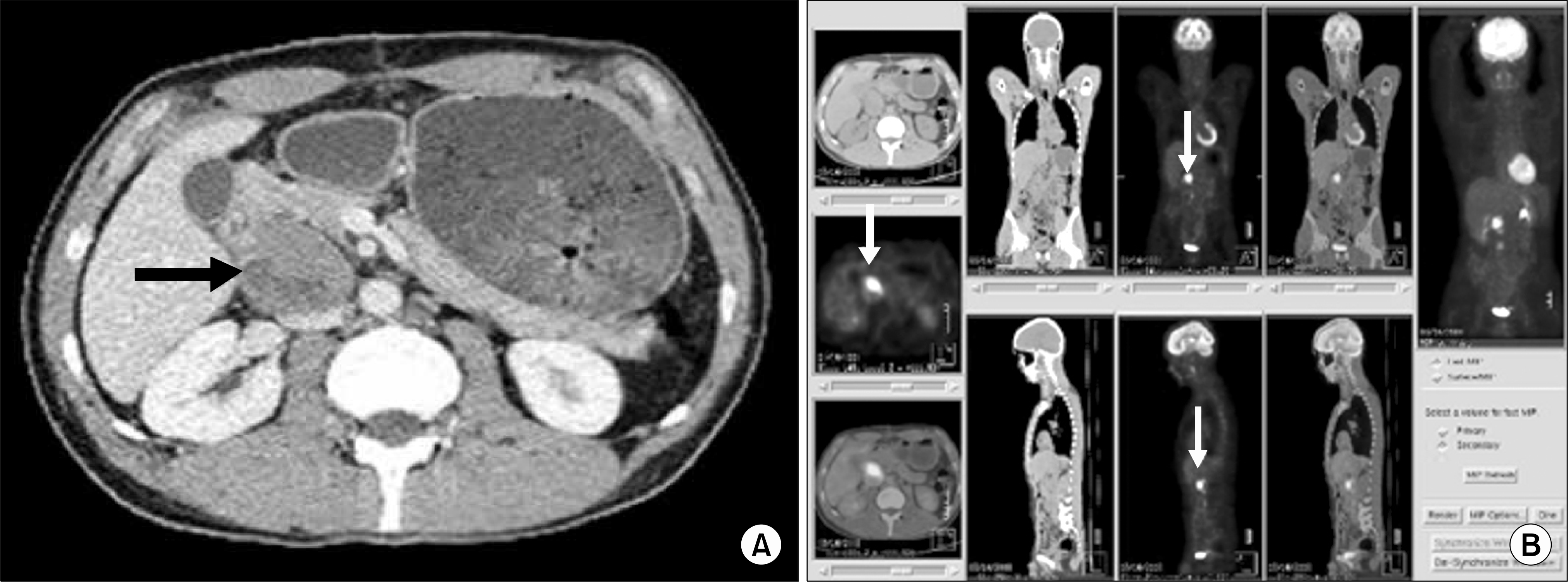
Fig. 2
Microscopic finding of a retroperitoneal mass shows seminoma cells with enlarged nucleoli in mitosis (black arrow) and many lymphocytes in the lobular septum (white arrow) (H&E, x400).
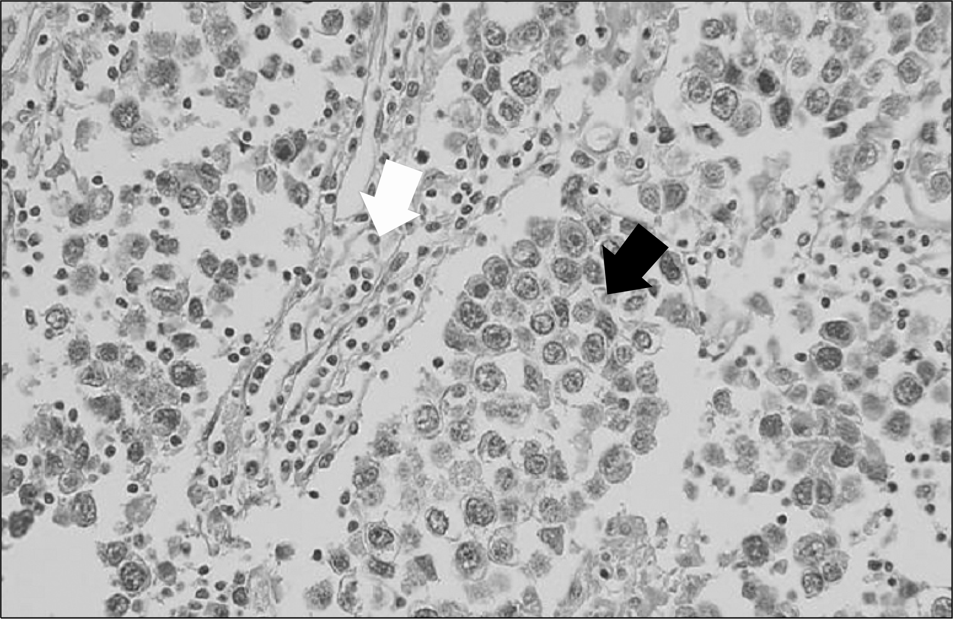
Fig. 3
(A) Ultrasonography of testis shows a segmental inhomogeneous lesion containing calcification in the atrophied right testis. (B) Magnetic resonance imaging (MRI) of testis shows a 33 mm ill-defined mass within the right testis that was not enhanced well and had heterogeneous low signal intensity on T2-weighted MRI.
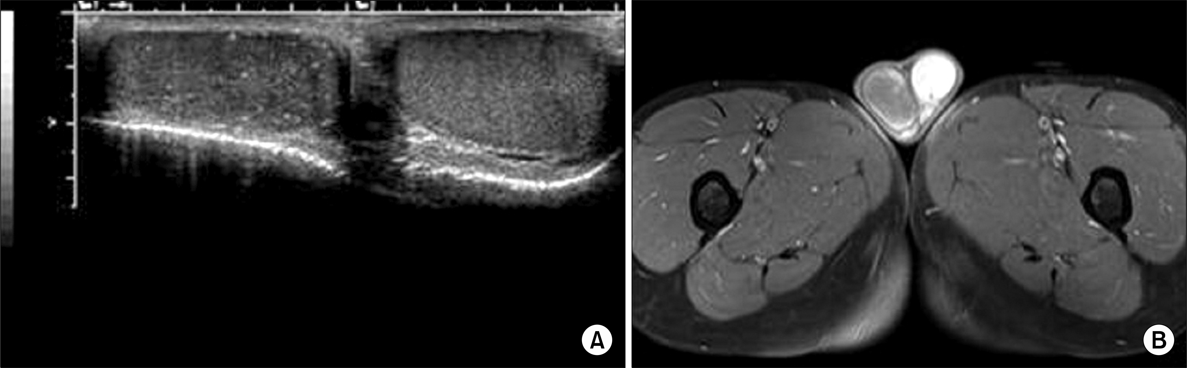
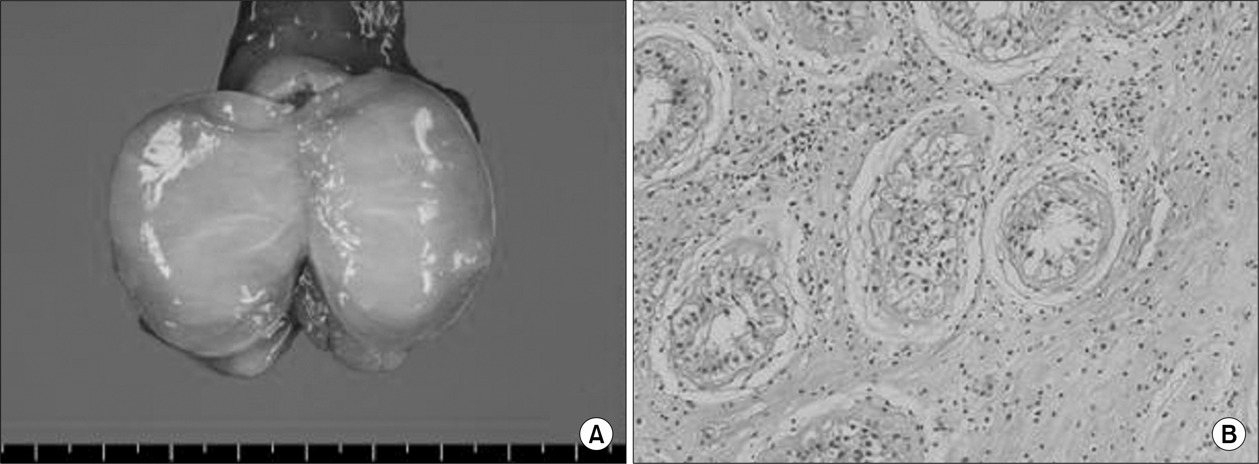
Fig. 4
(A) Gross finding of the right testis shows a well-defined, light yellow, solid scar-like mass, measuring 3.9x2.8 cm. The mass has replaced most of the testis and was abutting the tunica albuginea. (B) Microscopic finding of the right testis shows a tubular hyalinization dominant pattern with intratubular germ cell neoplasia (H&E, x400).
 XML Download