Abstract
The simultaneous occurrence of a renal cell carcinoma and a urothelial carcinoma in the same kidney is uncommon. Here we report the case of a 79-year-old woman with ipsilateral synchronous renal cell carcinoma and urothelial carcinoma. She was referred to our hospital for gross hematuria and right flank pain. A computed tomography scan showed a 15x20 mm enhanced lesion on the upper calyx and a 12x15 mm mass on the lateral aspect of the right kidney. We thus suspected a renal pelvis tumor and performed right hand assisted laparoscopic nephroureterectomy with bladder cuff excision (HALSNU). Gross findings were multiple, pale yellowish papillary masses on the upper and lower major calices, of which the largest one measured 16x20 mm. A separated solid mass measuring 12x16 mm was also noted on the anterior midportion of the kidney. The former was a urothelial carcinoma and the latter was a chromophobe renal cell carcinoma. We present a rare case of a chromophobe renal cell carcinoma and a urothelial carcinoma in the same kidney.
Go to : 
REFERENCES
1.Lee JW., Kim MJ., Song JH., Kim JH., Kim JM. Ipsilateral synchronous renal cell carcinoma and transitional cell carcinoma. J Korean Med Sci. 1994. 9:466–70.

2.Anseline P., Howarth VS. A case of transitional cell carcinoma of the renal pelvis, clear cell renal carcinoma, and analgesic nephropathy. Aust N Z J Surg. 1977. 47:521–3.

3.Michel K., Belldegrun A. Synchronous RCC and TCC of the kidney in a patient with multiple recurrent bladder tumors. Rev Urol. 1999. 1:99–103.
4.Terada T., Inatsuchi H., Yasuda M., Osamura Y. A kidney carcinoma with features of clear cell renal carcinoma and transitional cell carcinoma: a combined renal cell and transitional cell carcinoma? Virchows Arch. 2003. 443:583–5.

5.Hart AP., Brown R., Lechago J., Truong LD. Collision of transitional cell carcinoma and renal cell carcinoma. An immunohistochemical study and review of the literature. Cancer. 1994. 73:154–9.

6.Boring CC., Squires TS., Tong T., Montgomery S. Cancer statistics, 1994. CA Cancer J Clin. 1994. 44:7–26.

7.Janzen NK., Kim HL., Figlin RA., Belldegrun AS. Surveillance after radical or partial nephrectomy for localized renal cell carcinoma and management of recurrent disease. Urol Clin North Am. 2003. 30:843–52.

8.Crotty TB., Farrow GM., Lieber MM. Chromophobe cell renal carcinoma: clinicopathological features of 50 cases. J Urol. 1995. 154:964–7.

9.Han CH., Choi YJ., Sung SY., Kang SH., Yoon JY., Hwang TK, et al. Chromophobe cell renal carcinoma: a report of four cases with DNA ploidy and ultrastructural study. Korean J Urol. 1998. 39:91–6.
10.Steven CC., Andrew CN., Ronald MB. Renal tumors. Wein AJ, Kavoussi LR, Novick AC, Partin AW, Peters CA, editors. editors.Campbell-Walsh urology. 9th ed.Philadelphia: Saunders;2007. 1567-637.
Go to : 
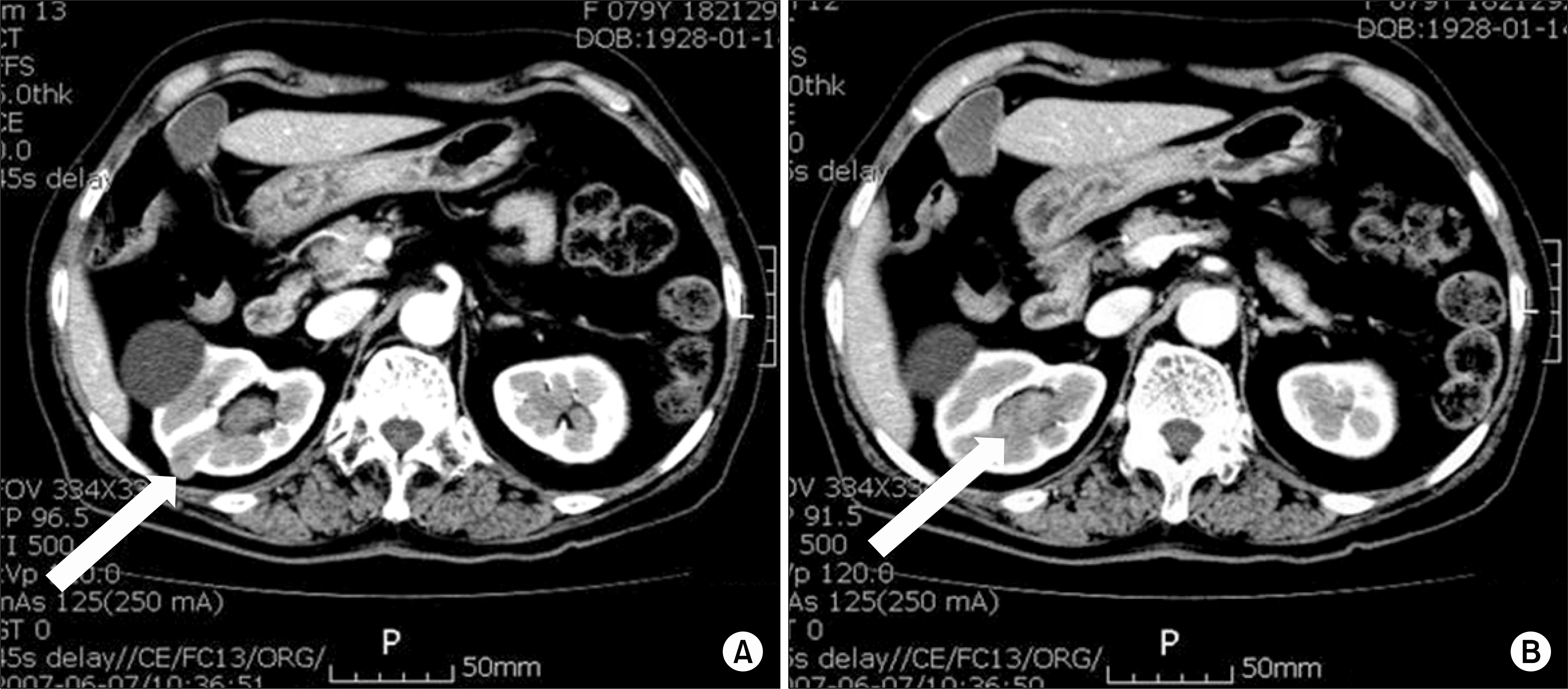
 | Fig. 1Abdominal CT scan showed a 12 mm mass on the lateral aspect, chromophobe renal cell carcinoma, and 15x20 mm mildly enhancing mass (arrow), urothelial carcinoma, on the upper calyx of the right kidney (B). |
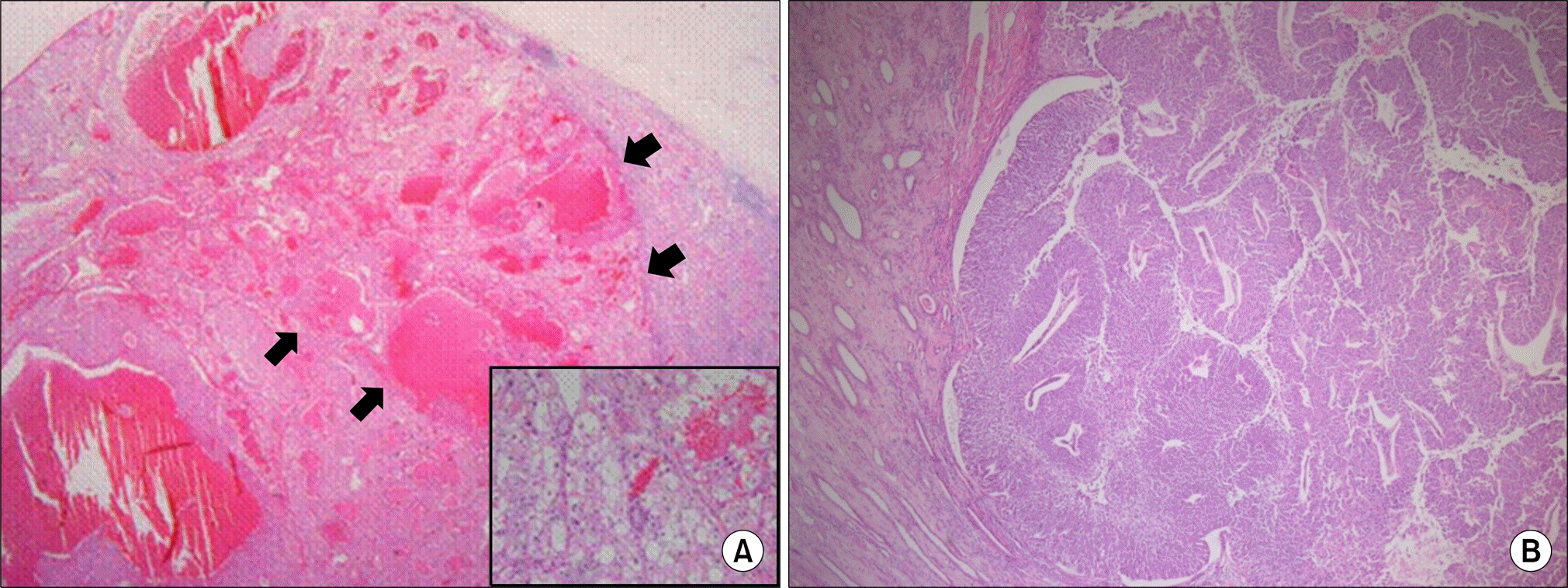
 | Fig. 2(A) A well-circumscribed solid mass (arrow) measuring 16x12 mm on the mid portion of the right kidney is noted (H&E, x12.5) (Inlet) The tumor cells revealed a high nuclear grade (Fuhrman nuclear grade 4) of chromophobe renal cell carcinoma (H&E, x200) (B) Multiple papillary urothelial carcinomas, high grade, are found on the upper and lower calyx, which were not invading the subepithelium (H&E, x40). |


 PDF
PDF ePub
ePub Citation
 Print
Print


 XML Download