

 PDF
PDF ePub
ePub Citation
 Print
Print


Pheochromocytoma is a rare neoplasm of the chromaffin tissue. Related tumors in extra-adrenal sympathetic and parasympathetic paraganglia are classified as extra-adrenal paragangliomas. A pheochromocytoma is an intra-adrenal sympathetic paraganglioma. Approximately 220 cases have been reported in the literature. These tumors account for less than 0.06% of all urinary bladder tumors.1 Females are affected more frequently than males, and the tumors are more common between the second and fourth decades of life.1 Paragangliomas are generally benign tumors, with only less than 10% being malignant. Most of these tumors are hormonally active and secrete mainly noradrenaline (rarely adrenaline), calcitonin, and adrenocorticotropic hormone. The clinical symptoms of the tumors are related to these hormonal secretions. Here we report a case of a paraganglioma arising in a urinary bladder with a bladder stone.
CASE REPORT
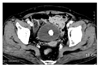
A 79-year-old woman presented with intermittent, painless gross hematuria for 7 days. Intermittent lower abdominal pain was also seen. She reported 2 past medical histories of 3 to 4 occurrences of recurrent cystitis about 4 to 5 years ago and high blood pressure. No significant abnormalities were found on the physical examination. The results of a cell blood count and routine blood chemistry tests, including cell cytologies that were repeated 3 times, were within normal limits. Urinalysis, however, showed RBC: many/HPF. A pelvic CT scan from a local medical center demonstrated a 2×2 cm calcification in the posterior wall of the bladder (Fig. 1), but no mass-like lesion was seen in the bladder. Cystoscopy showed a 2×2 cm brown stone in the posterior wall of the bladder; it was fragmented and removed. After removal of the bladder stone, a 1.5×2.0 cm bulbous and erythematous intravesical mass with no stalk was observed separately posterior to the stone and was removed completely with a cystoscope. No elevation of blood pressure was seen while removing the mass. The patient's hematuria resolved 3 days after vesicolithotripsy and transurethral resection of the bladder mass, so we removed the Foley catheter.
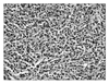
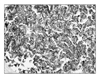
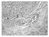
Pathologic examination of the bladder mass showed that most of the tissue taken from the biopsy was substituted by malignant cells composed of nests of cuboidal epithelial cells surrounding a delicate vasculature. The classic "zellballen" pattern in an adrenal tumor showed architectural variations. The tumor cells were characterized by relatively uniform round nuclei with fine chromatin and abundant amphophilic cytoplasm (Fig. 2). We also observed calcification within malignant tissue but no relationship with the stone was found. In immunohistochemical staining for CD56 (Zymed, USA), chromogranin (DAKO, Denmark), and nonspecific enolase (DAKO), the tumor cells showed a diffuse, strong, positive reaction to CD56, chromogranin, and nonspecific enolase. By contrast, the tumor cells showed a negative reaction to cytokeratin 7 (CK 7; Neomarkers, USA), CK 20 (Neomarkers), and epithelial membrane antigen (DAKO). These results confirmed that the specimen was composed of neuro-endocrinologic tumor cells, and we diagnosed a paraganglioma in the bladder (Fig. 3) and the tumor infiltrates into the proper muscular layer (Fig. 4). We recommended a partial cystectomy according to the biopsy, but the patient refused our recommendation. Thus, only repeated cystoscopic exams were performed 3 months and 6 months after the surgery in our outpatient department. Additional biopsies showed only chronic inflammatory changes from postoperative scarring in repeated cystoscopic exams.
DISCUSSION
The urinary bladder pheochromocytoma was similar to an adrenal pheochromocytoma and consisted of chromaffin tissue from the neural crest embryologically. Zimmerman et al described a pheochromocytoma for the first time in 1953,1 and an additional 220 cases have been reported since then, including in Korea.2 Paragangliomas account for 10% of total pheochromocytomas, but a paraganglioma in the bladder is extremely rare. This is because of poor degeneration of chromaffin cells including its neural system, whereas the sympathetic system is distributed throughout the entire bladder layer.3
Most pheochromocytomas in the bladder are hormonally active (e.g., noradrenaline, calcitonin, and adrenocorticotropic hormone) and are mostly asymptomatic, causing paroxysms of hypertension, syncope, tachycardia, and headache on filling or emptying of the bladder. Headache with urination or palpitation, visual symptoms, or diaphoresis were shown in 47% to 77% of patients, and similar symptoms occur in defecation and ejaculation.4 The most common symptom of paraganglioma is hypertension, which occurs in 65% to 75% of paraganglioma patients. Hematuria occurs in 55% to 58%.5-7 In patients with gross hematuria and hypertension, checking for an elevation of catecholamine in a 24-hour urine collection might be helpful for preoperative diagnosis.8 However, some patients whose catecholamine level in urine is normal would present only hypertension while voiding. These are the reactions from the neuropeptide Y, somatostatin, vasoactive intestinal peptide, gastrin, and serotonin secreted by the tumor.8 Patients with a paraganglioma in the bladder show a decreased level of converting enzyme, which converts norepinephrine to epinephrine. But the level of secreted norepinephrine while urinating would be elevated by chromaffin cells. Thus, the comparison of norepinephrine levels before urination and 3 hours after urination has diagnostic value.8 In the present case, preoperative computed tomography showed no intravesical mass and only calcification. We did not proceed with these analyses because we thought the lesion was a vesical stone. For those who present with hematuria with hypertension, we recommend considering a paraganglioma.
The pathological findings of paraganglioma are typically nests of cuboidal epithelial cells surrounding a delicate vasculature. The classic "zellballen" pattern in an adrenal tumor shows architectural variations. Tumor cells are characterized by relatively uniform round nuclei with fine chromatin and abundant amphophilic cytoplasm.9 Circular calcification is rarely found,10 but in this case, microscopic calcification is one and vesical stone is another. It is not easy to distinguish between paragangliomas with muscular invasion and urothelial carcinomas because of their similar features. With immunohistochemical staining, paragangliomas are positive for neuroendocrine markers and negative for cytoskeletal antigen, whereas urothelial tumors are negative and positive, respectively.9 This also supports our case being a neuroendocrine tumor because it expressed diffuse strong positive for CD56, chromogranin, and nonspecific enolase and negative for CK 7, CK 20, and epithelial membrane antigen.
The most effective management for a paraganglioma is surgical resection.8 It was reported that in most cases, once a paraganglioma is diagnosed pathologically, then partial cystectomy is the treatment of choice rather than a transurethral resection of the bladder. There are several reasons for this. Transurethral resection can irrigate the tumors and induce the blood pressure to fluctuate, which increases the risk, and it is very difficult to control its depth and extent. Pheochromocytomas can easily recur if the tumor cannot be resected completely. It is important to resect the main tumor and adjacent tissue completely because of the high tendency for malignancy compared with paragangliomas derived from the same neural crest, although more than 90% of tumors seem to present with benign features.6 In the present case, because the tumor infiltrated the proper muscular layer, we recommended a partial cystectomy, but the patient refused our recommendation. Thus, only repeated cystoscopic exams were performed 3 months and 6 months after the operation, and no evidence of recurrence was seen. If localized invasion or enlargement of the lymph nodes is detected, a radical cystectomy with lymph nodes dissection should be considered although distant long metastasis to enhance the survival rate. The effectiveness of chemoradiation therapy for pheochromocytomas is limited. Palliative radiation therapy can be considered in patients with multiple painful bony metastases or with symptoms of spinal cord compression.
 XML Download