

 PDF
PDF ePub
ePub Citation
 Print
Print


Abstract
Spontaneous and post-traumatic renal intracystic hemorrhages are extremely rare, but are a potential danger to patients with cystic kidney disease. We report two cases of post-traumatic intracystic massive hemorrhage in renal cysts. One patient was a 27-year-old male who presented with left flank pain and gross hematuria after slipping on the stairs 2 days previously. The other patient was a 58-year-old male who presented with back pain due to an accident. The circulatory states of the two patients were deteriorated and renal intracystic hemorrhages were detected on computed tomography. One patient underwent a simple nephrectomy and the other patient was treated with arterial embolization. We present two cases of renal intracystic hemorrhage, emphasizing early diagnosis and the treatment of choice.
Go to : 
References
1. McLaughlin AP 3rd, Pfister RC. Spontaneous rupture of renal cysts into the pyelocaliceal system. J Urol. 1975; 113:2–7.

2. Hughes CR, Stewart PF Jr, Breckenridge JW. Renal cyst rupture following blunt abdominal trauma: case report. J Trauma. 1995; 38:28–9.
3. Rauschmeier H, Jakse G. Traumatic rupture of renal cyst into the retroperitoneum and the pyelocalyceal system. Int Urol Nephrol. 1983; 15:117–9.
4. Papanicolaou N, Pfister RC, Yoder IC. Spontaneous and traumatic rupture of renal cysts: diagnosis and outcome. Radiology. 1986; 160:99–103.
5. Rainio J, De Giorgio F, Carbone A. Death from renal cyst: spontaneous or traumatic rupture? Am J Forensic Med Pathol. 2006; 27:193–5.
6. Redmond PL, Royle G. Rupture of a calcified renal cyst following blunt abdominal trauma. Br J Radiol. 1985; 58:683–4.
7. Lopez Cubillana P, Hita Rosino E, Asensio Egea L, Rigabert Montiel M, Hita Villaplana G, Perez Albacete M. Wunderich syndrome. Review of its diagnosis and therapy. Report of seven cases. Actas Urol Esp. 1995; 19:772–6.
8. Hwang HH, Cheon SH, Moon KH, Lee SK, Choo HS, Hwang JC, et al. Renal ruptures with active bleeding treated with emergency selective renal arterial embolizaton. Korean J Urol. 2008; 49:177–81.
Go to :
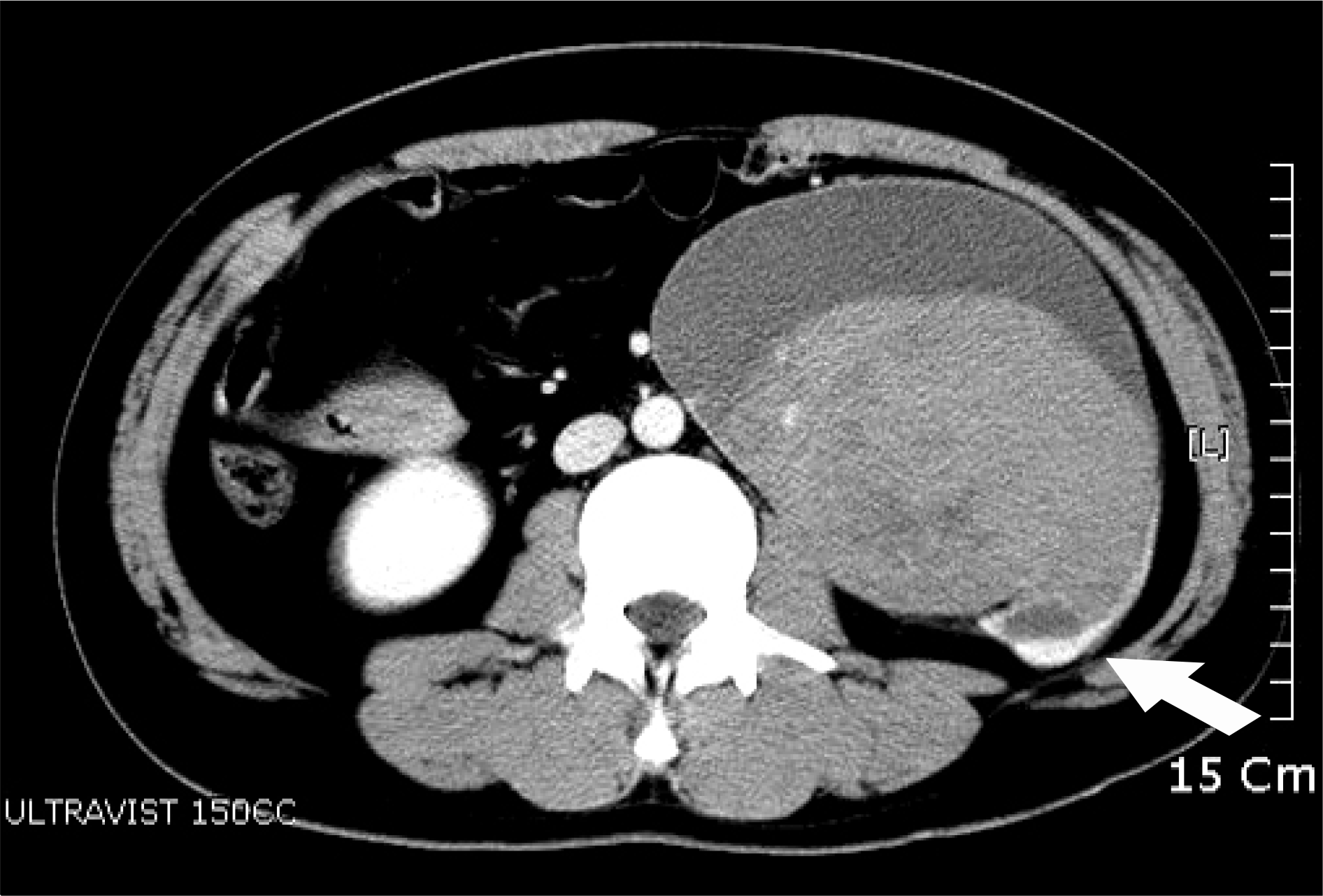 | Fig. 1.Abdominal computed tomography shows a large left retroperitoneal mass with a large hematoma and thinning of the left lower pole of the parenchyma (arrow). |
 | Fig. 2.The kidney measures 21x17x13cm and weighs 2,130g. It is dilated cystically and filled with blood and blood clots. (A) The renal parenchyma is thinned markedly, measuring <0.1cm in thickness. (B) The cysts are lined by urothelial cells and several immature tubules are present in the remaining renal parenchyma (H&E, x100). |
 | Fig. 3.(A) and (B) show active contrast extravasation in the left renal cyst (white arrow) and intracystic hematoma formation (initial). (C) and (D) show persistent active contrast extravasation in the left renal cyst and increasing intracystic and retroperitoneal hematoma formation (thin arrow; 6cm (B) to 12cm (D)) (12 hours later). |
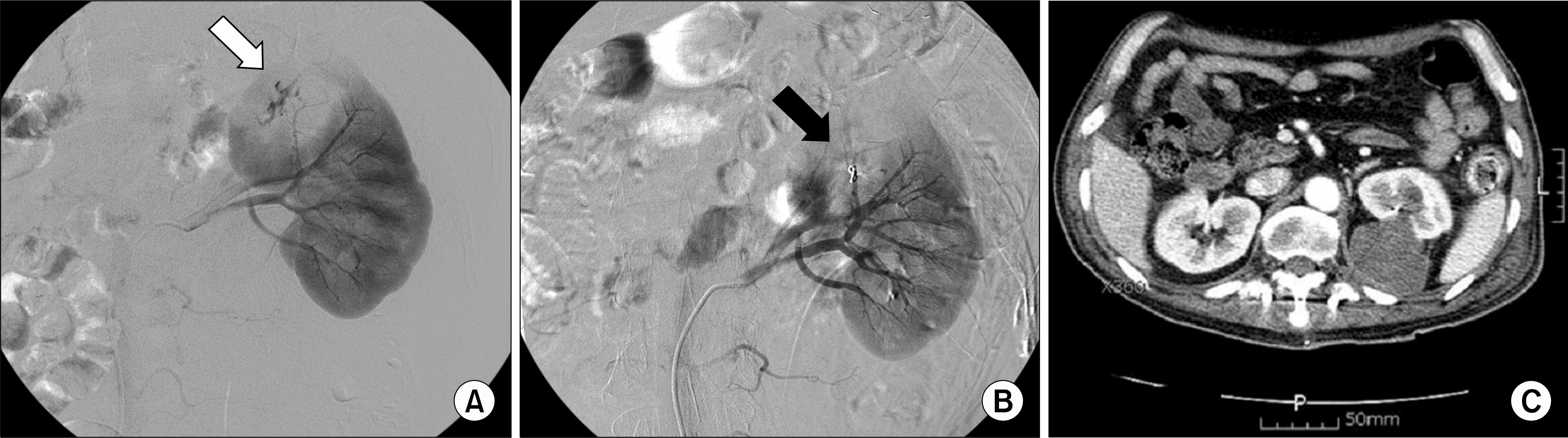 | Fig. 4.(A) Angiography reveals active extravasation from the segmental branch of the left renal artery (white arrow). (B) There is no extravasation after selective embolization using gelfoam and coils (black arrow). (C) No evidence of contrast extravasation and more absorbing state of previously noted intracystic hematoma on the follow-up CT scan (3 weeks later). |
 XML Download