

 PDF
PDF ePub
ePub Citation
 Print
Print


Abstract
Purpose
We assessed the impact of using a porcine model on the training of laparoscopic partial nephrectomy (LPN) and compared the training effectiveness between surgeons with and without previous laparoscopic experience.
Materials and Methods
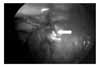
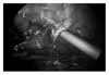
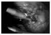
Surgeon A had previous laparoscopic experience, with the exception of LPN, while surgeon B had no prior laparoscopic experience. A tumor model was created by subcapsular injection of liquid plastic (Smooth-Cast 320) in the kidney. We recorded the total operation time, the bowel dissection time, the renal pedicle dissection time, the warm ischemic time, the mass resection time, the suture time, and the presence of major complications for each surgeon.
Results
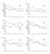
The mean operation time was significantly shorter for surgeon A compared to surgeon B (49.1±4.5 and 63.6±8.4 minutes, respectively, p< 0.001). Although the mass resection time was significantly shorter for surgeon A as well, there were no significant differences between the two surgeons in terms of warm ischemia time and suture time. As the training progressed, surgeon B improved in all surgical steps and surgeon A showed improvement in time for warm ischemia and suturing the defect. Five complications occurred (two cases by surgeon A and three cases by surgeon B).
Figures and Tables

References
1. Clayman RV, Kavoussi LR, Soper NJ, Dierks SM, Meretyk S, Darcy MD, et al. Laparoscopic nephrectomy: initial case report. J Urol. 1991. 146:278–282.
2. Traxer O, Gettman MT, Napper CA, Scott DJ, Jones DB, Roehrborn CG, et al. The impact of intense laparoscopic skills training on the operative performance of urology residents. J Urol. 2001. 166:1658–1661.
3. Soper NJ, Hunter JG. Suturing and knot tying in laparoscopy. Surg Clin North Am. 1992. 72:1139–1152.
4. Wolfe BM, Szabo Z, Moran ME, Chan P, Hunter JG. Training for minimally invasive surgery. Need for surgical skills. Surg Endosc. 1993. 7:93–95.
5. Medina M. Formidable challenges to teaching advanced laparoscopic skills. JSLS. 2001. 5:153–158.
6. Chandrasekera SK, Donohue JF, Orley D, Barber NJ, Shah N, Bishai PM, et al. Basic laparoscopic surgical training: examination of a low-cost alternative. Eur Urol. 2006. 50:1285–1290.
7. Breda G, Nakada SY, Rassweiler JJ. Future developments and perspectives in laparoscopy. Eur Urol. 2001. 40:84–91.
8. Corvin S, Oberneder R, Adam C, Frimberger D, Zaak D, Siebels M, et al. Use of hydro-jet cutting for laparoscopic partial nephrectomy in a porcine model. Urology. 2001. 58:1070–1073.
9. Katz R, Hoznek A, Antiphon P, Van Velthoven R, Delmas V, Abbou CC. Cadaveric versus porcine models in urological laparoscopic training. Urol Int. 2003. 71:310–315.
10. Hidalgo J, Belani J, Maxwell K, Lieber D, Talcott M, Baron P, et al. Development of exophytic tumor model for laparoscopic partial nephrectomy: technique and initial experience. Urology. 2005. 65:872–876.
11. Fried GM, Derossis AM, Bothwell J, Sigman HH. Comparison of laparoscopic performance in vivo with performance measured in a laparoscopic simulator. Surg Endosc. 1999. 13:1077–1081.
 XML Download