Female urinary retention is extremely rare. Two cases of female urethral adenocarcinoma that presented as urinary retention are reviewed and discussed.
The incidence of primary urethral adenocarcinoma in females is less than 1%. In patients with urinary retention or obstructive voiding symptoms, an increased index of suspicion for urethral adenocarcinoma is imperative in order to provide early aggressive therapy for this malignancy. We report 2 cases of female urethral adenocarcinoma presenting with urinary retention.
CASE REPORTS
CASE 1
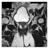
A 62-year-old woman presented with symptoms of decreased force of stream, and two times acute urinary retention about 580 ml, 620 ml. She was treated with a urethral catheterization to relieve an acute micturition pain and spontaneous urinary retention. She had no history of hematuria or urinary disturbance, but only hypertension. Pelvic examination revealed a stony hard mass around urethra with no other abnormalities at vagina. On blood chemistry, only serum creatinine was slightly increased (1.7 mg/dl). And urinalysis was normal finding. Urodynamic study confirmed bladder outlet obstruction with no detrusor instability. Evaluation revealed bladder neck mass with bilateral hydronephrosis on ultrasonography. Cystoscopy confirmed a midurethral stony hard mass at the 12-3 o'clock position and transurethral biopsy was done. The pathology report revealed a urethral adenocarcinoma. For more detail information, pelvic magnetic resonance imaging study (MRI) was done. MRI showed about 3.3×2.9 cm sized well enhancement urethral mass with surrounding cystic component such as urethral cancer from urethral diverticulum (Fig. 1). No demonstrable lymph nodes enlargement in pelvic cavity. Gastrointestinal and gynecologic evaluation revealed no other primary source for the malignancy. The patient underwent pelvic exenteration and ileal conduit. On gross histopathologic examination, the tumor was largely confined to the periurethral region (Fig. 2). On microscopic histopathologic examination, it infiltrated into the subepithelial stroma of the urethra and the bladder neck. It was a moderately differentiated mucinous adenocarcinoma (Fig. 3). There was no definite lymphovascular permeation. All the resection margins were clear. Lymph nodes were identified in the lymphadenectomy specimen and were free of metastatic disease. This patient still alive in good health for 17 months after operation.
CASE 2
A 64-year-old woman was referred for evaluation of newly onset urinary retention of unclear etiology. She had history of dysuria, voiding difficulty and urinary incontinence for 3 months and suffered from both flank pain. Serum creatinine was 2.1 mg/dl. Urethral catheterization was tried to relieve an acute urinary retention but not passed. And then, suprapubic cystostomy was done. Urodynamic study revealed a high pressure, low flow voiding pattern with a postvoid residual volume of 500 ml, consistent with bladder outlet obstruction. Ultrasonography showed both moderate hydronephrosis, severe trabeculation of bladder and pelvic mass lesion. Pelvic examination and palpation of the anterior vaginal wall confirmed a firm mass lateral to the left proximal urethra. On cystourethroscopy with a 19.5F cystoscopy, it was difficult to pass into the urethra, Irregular urethral mucosa on the left side of the proximal urethra was biopsied transurethrally with cold-cup biopsy forceps. Pathologic examination revealed poorly differentiated adenocarcinoma. After serum creatinine was normal level, for further evaluation of pelvic mass, computed tomography (CT) was done. CT showed ill-defined low attenuated mass at bladder neck with bilateral hydronephrosis. Gastrointestinal and gynecologic evaluation revealed no other primary source for the malignancy. Operation was recommended. But she refused and not followed up.
DISCUSSION
Primary female urethral cancer comprises less than 1% of all malignancies.1
Adenocarcinoma is the most common histopathology detected within female urethral diverticula (61%), with only 12% showing squamous cell carcinoma despite the fact that the majority of the urethra is lined with squamous epithelium. The most common histopathology in nondiverticular urethral malignancy is squamous cell carcinoma.2
Our histopathology was adenocarcinoma with diverticulum.
There are two major histologic subtypes for female urethral adenocarcinoma: columnar/mucinous and clear cell adenocarcinoma.3 If cause of urinary retention was unclear etiology, especially in patients with no neurologic symptoms, no prior pelvic surgery, and no pelvic prolapse, cystourethroscopy should be performed, at which time the urethra is palpated over the cystoscopic sheath through the anterior vaginal wall. It is important to find of the suspicion of malignancy. Transurethral biopsies with cold-cup biopsy forceps and transvaginal needle biopsies of the urethral mass should be performed promptly to expedite early treatment of this aggressive malignancy.
The prognosis of female urethral tumor depends on the primary site and tumor size and stage, but not on histo1ogic type and grade. The 5-year survival rate of carcinoma of the proximal urethra is approximately 10%.4
There is no consensus on the treatment of women with urethral cancer. This lack of consensus is due to the rarity of the tumor and the poor prognosis of women with large lesions irrespective of their treatment. Women with small lesions in the distal urethra are treated with surgery or radiotherapy. Large tumors and tumors of the proximal urethra are treated with radiotherapy in an attempt to preserve anatomy and function of the urethra, bladder, and vagina. Very large lesions (i.e., greater than 4 cm) are treated with combinations of surgery, radiotherapy, and chemotherapy.5 Mayer et al6 performed radical surgery on ten women with advanced urethral cancer. All women had a radical cystectomy with anterior vaginectomy and total urethrectomy. Of these ten women only three were free of disease with limited follow-up.
In 86% of the cases of urethral adenocarcinoma treated with radiation therapy, and in 41% of the cases, the patients underwent an anterior exenteration or a cystectomy.
However, despite aggressive therapy for urethral carcinoma, recurrence rates are high and the optimal course of therapy has not been determined.7
Bladder outlet obstruction in women with no neurologic complaints, no history of pelvic surgery, and no pelvic prolapse should raise the suspicion of urethral malignancy. Urethral malignancy must always be included in the differential diagnosis of any female patient with obstructive symptoms. High clinical suspicion and accurate preoperative diagnosis will result in the proper aggressive therapy necessary in treatment of this rare disease.


 PDF
PDF ePub
ePub Citation
 Print
Print


 XML Download