

 PDF
PDF ePub
ePub Citation
 Print
Print


Abstract
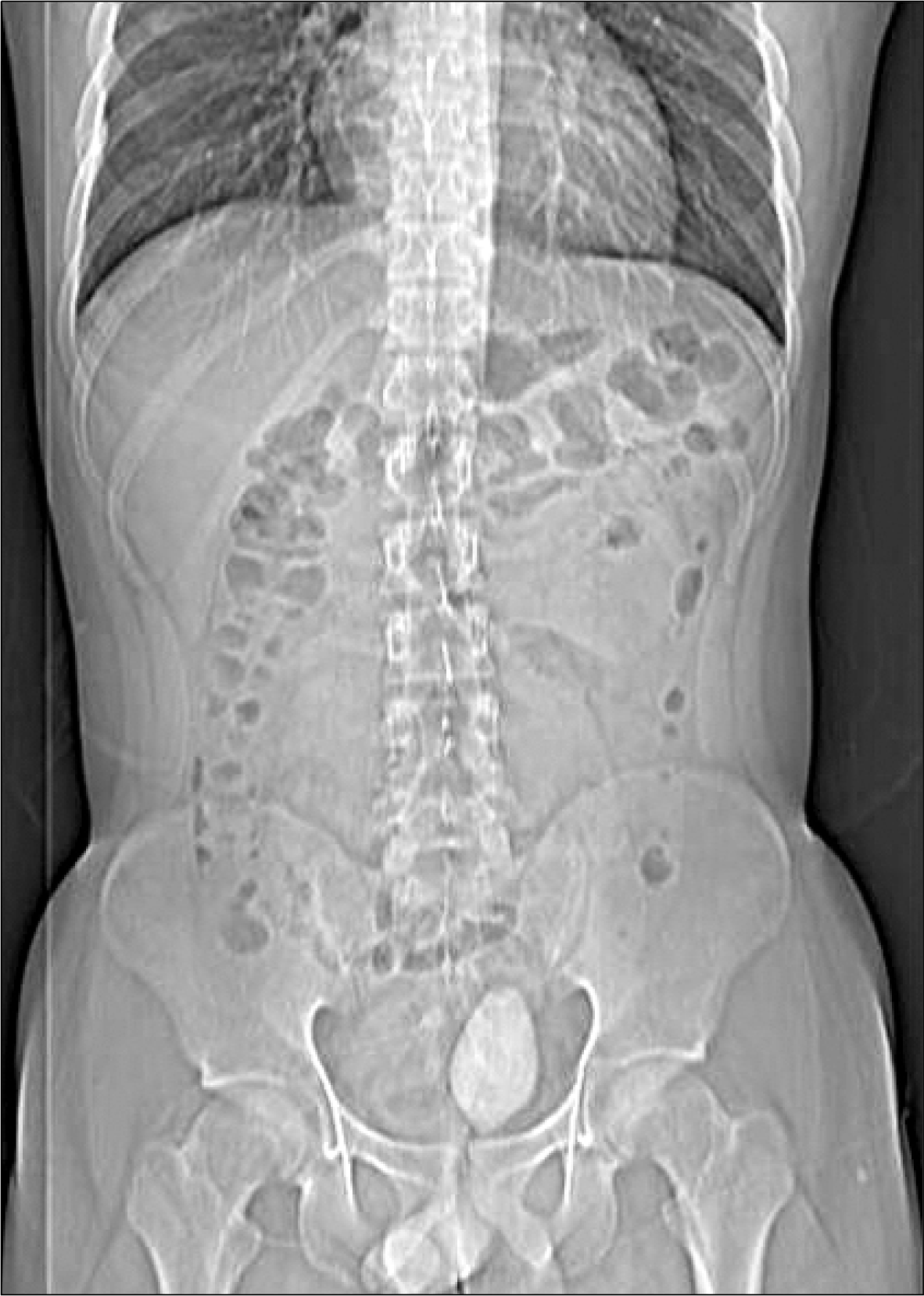
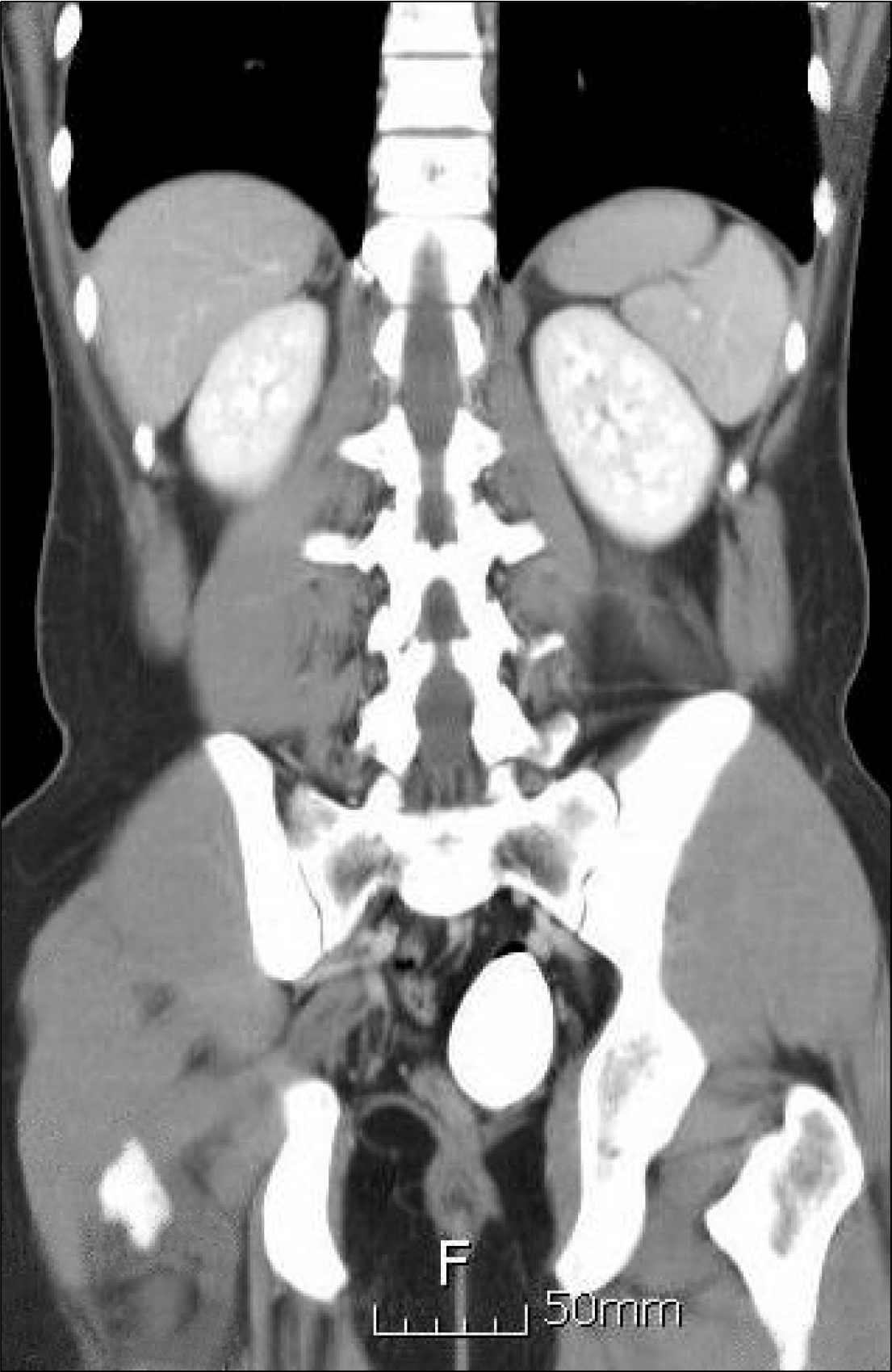
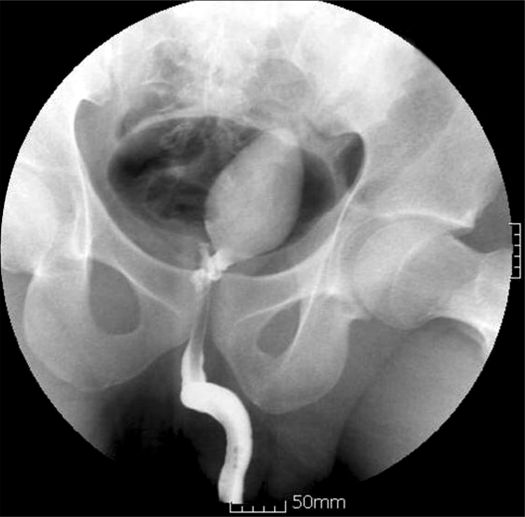
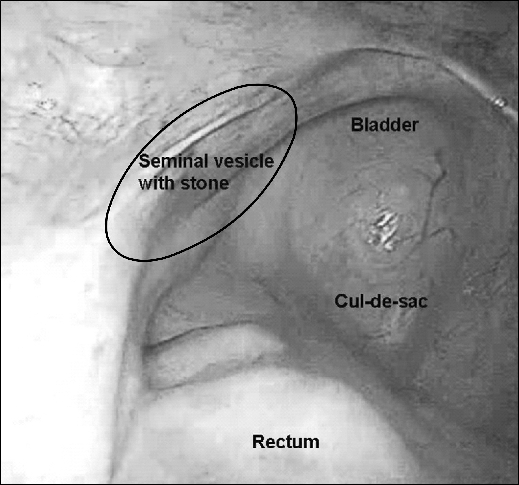
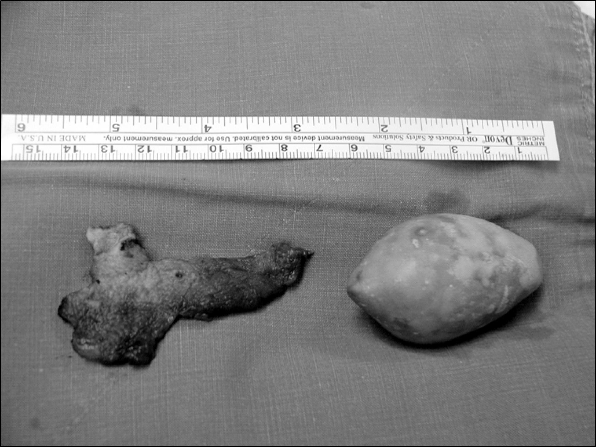
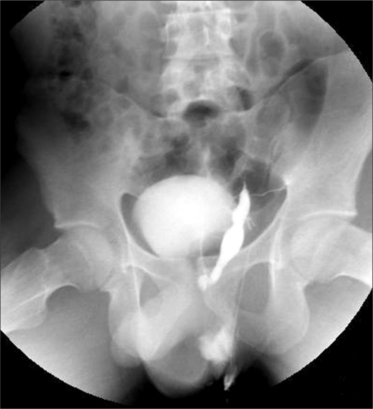
Stones in the seminal vesicle are extremely rare. We report a case with a large stone in a dilated seminal vesicle. A 20-year-old man presented with a large calcified density in the pelvic cavity on plain films. A 6.0 cm cone shaped stone was noted in the dilated left seminal vesicle diagnosed by radiological examination. We treated the patient by transperitoneal laparoscopic stone removal and partial seminal vesiculectomy. The composition of stone was carbonate apatite. This approach to the treatment of such pathological conditions of the seminal vesicles provides an additional option.
REFERENCES
1.Amano T., Kunimi K., Ohkawa M. Transrectal ultrasonography of the prostate and seminal vesicles with hemospermia. Urol Int. 1994. 53:139–42.

2.Sandlow JI., Winfield HN., Goldstain M. Surgery of the scrotum and seminal vesicles. Wein AJ, Kavoussi LR, Novick AC, Partin AW, Peters CA, editors. editors.Campbell-Walsh urology. 9th ed.Philadelphia: Saunders;2007. 1098-127.
3.Modi PR. Case report: endoscopic management of seminal vesicle stones with cutaneous fistula. J Endourol. 2006. 20:432–5.
4.Cuda SP., Brand TC., Thibault GP., Stack RS. Case report: endoscopic laser lithotripsy of seminal-vesicle stones. J Endo-urol. 2006. 20:916–8.
5.Uchijima Y., Hiraga S., Akutsu M., Yoshida K., Hobo M., Okada K. Stones of the seminal vesicles and ejaculatory duct in infant: report of a case. Hinyokika Kiyo. 1984. 30:1843–9.
6.Carachi R., Gobara D. Recurrent epididymo-orchitis in a child secondary to a stone in the seminal vesicle. Br J Urol. 1997. 79:997.
7.Li YK. Diagnosis and management of large seminal vesicle stones. Br J Urol. 1991. 68:322–3.
8.Wesson L., Steinhardt G. Case profile: seminal vesicle stones. Urology. 1983. 22:204–5.
9.Orquiza CS., Bhayani BN., Berry JL., Dahlen CP. Ectopic opening of the ureter into the seminal vesicle: report of case. J Urol. 1970. 104:532–5.
10.Yang SC., Rha KH., Byon SK., Kim JH. Transutricular seminal vesiculoscopy. J Endourol. 2002. 16:343–5.
 XML Download