

 PDF
PDF ePub
ePub Citation
 Print
Print


Lymphoepithelioma is a malignant epithelial tumor that was originally described within the nasopharynx.1 Tumors with a similar histology have been described in other organs; salivary gland, thymus, cervix, skin, lung, stomach, and bladder.
Lymphoepithelioma-Like Carcinoma of the urinary system is rare. We report a case of Lymphoepithelioma-like Carcinoma confined to the renal pelvis.
CASE REPORT
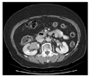
A 70-year-old woman presented with microscopic hematuria and right hydronphrosis. Excretory urography showed medial displacement of right renal pelvis with a filling defect that had an irregular contour, and dilatation of right upper and interpolar calyces. Cystoscopy revealed no abnormal finding in the urinary bladder. Computerized tomography (CT) of the abdomen and pelvis demonstrated a 4×3 cm tumor in the dilated right renal pelvis and extension of the tumor to anterior calyces (Fig. 1). Also there was no evidence of lymph nodes metastasis and invasion of renal parenchyma on CT. Chest x-ray did not reveal metastatic disease.
Nephroureterectomy with bladder cuff excision was performed. A 4.2×3.9×2.8 cm, well circumscribed polypoid mass was noted in the major calyx and pelvis of lower pole of the right kidney. The color and consistency of the cut surface of the mass was gray and firm, respectively. The mucosa of remaining calyx and ureter was grossly unremarkable.
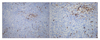
Microscopic examination of the resected tumor revealed a high-grade urothelial carcinoma, which had sheets of large tumor cells with a loss of cellular polarity. The cancer cells had marked nuclear pleomorphism and prominent nucleoli with a high nuclear-cytoplasm ratio. The stroma demonstrated marked infiltration of lymphoplasma cells. There was invasion of muscularis by tumor but neither lymphovascular emboli or involvement of renal parenchyma and ureter. Resection margin was free of tumor cells. Immunohistochemistry revealed the tumor cells to express pancytokeratin, cytokeratin 20 and epithelial membrane antigen. The tumor cells are weakly focal positive for cytokeratin 7 and are negative for vimentin. The infiltrated lymphocytes were composed of abundant CD3 positive T cells (Fig. 2) and a few CD20 positive B cells (Fig. 3). In situ hybridization for Epstein-Barr virus was negative. Based on the finding of morphological feature and immunostaining, we concluded that the diagnosis was lymphoepithelioma-like carcinoma of the renal pelvis. The stage of cancer is T2N0M0.
DISCUSSION
There are a few case reports of lymphoepithelioma-like carcinoma of the renal parenchyma and the urinary tract such as renal pelvis, ureter, and bladder.2 The most common involved site among urinary tract is the urinary bladder.3
Owing to the prominent inflammatory background, lymphoepithelioma-like carcinoma can be misdiagnosed either as a reactive inflammatory lesion or lymphoma.4 Immunochemistry for keratins and lymphoid markers can resolve this differential diagnosis.5
There has been not yet well known about the prognosis of Lymphoepithelioma-Like Carcinoma of the urinary system. Of these factors that affect the prognosis, the proportion of component of lymphoepithelioma-like carcinoma, that is, whether it is pure type or mixed type, is important and pure type tends to be more favorable outcome than mixed type.2 Our case is mixed type.
In case of muscle invasive bladder cancer, compared with cancer of different histological type, lymphoepithelioma-like carcinoma of the bladder has a better prognosis than others when treated with combination chemotherapy.6 Dinney et al6 reported three cases of muscle invasive lymphoepithelioam-like carcinoma of the bladder that was treated with combination chemotherapy. Two of three survived more than 5 years with no evidence of recurrence or metastasis.
Because we performed radical nephroureterectomy with bladder cuff excision and the tumor was confined to renal pelvis, the patient received no adjuvant treatment after surgery. We performed follow-up chest PA, abdomen and pelvic CT at 6, 12, 24 months and showed no evidence of recurrence or distant metastasis. The patient is still alive.

 XML Download