Abstract
Purpose
In the present study, we identified the pre-operative predictive factors of insignificant prostate cancer and we analyzed their diagnostic accuracy.
Material and Methods
Of a total 343 patients who had undergone radical prostatectomy, 33 patients (9.6%) were diagnosed with insignificant cancer that was characterized by a total cancer volume ≤0.5cc, a Gleason score (GS)≤6, a T stage≤2c and no positive surgical margin. We found the statistically significant factors after comparing of preoperative clinicopathological findings between two groups and determined the diagnostic accuracy of the identified predictors.
Results
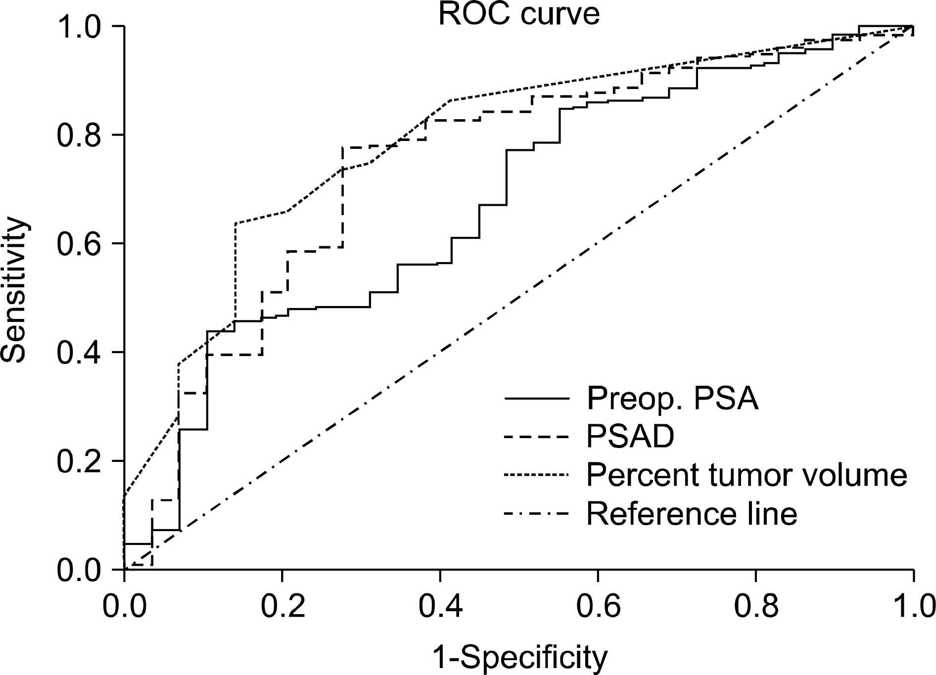
Of several factors, prostate-specific antigen (PSA) level (p=0.04, odds ratio (OR)=4.3 3.589≤95%confidence interval (CI)≤5.692), PSA density (PSAD) (p=0.01, OR=6.6, 2.115≤95%CI≤278.826), biopsy GS (p=0.03, OR=4.6, 1.114≤95%CI≤12.568) and volume of the largest cancer (p=0.02, OR=5.6, 2.471≤95%CI≤9.725) were analyzed as independent predictors of insignificant cancer. The volume of the largest cancer was the most precise predictor (AUC=0.791), followed by the PSAD (AUC=0.748) and the PSA level (AUC=0.677) in the ROC (receiver operating characteristic) curve analysis. The sensitivity, specificity and positive predictive value for predicting insignificant cancer were 10.3%, 63.7% and 12.8% at a PSA level of 10ng/ml, and 44.8%, 16.8% and 26.3% at a PSAD of 0.15ng/ml/ml, and 13.8%, 53.8% and 14.2% at a volume of the largest cancer of 50%, respectively. Even with using a combination of these three factors as well as a biopsy GS≤6, only 53% of insignificant prostate cancer could be predicted preoperatively.
Go to : 
REFERENCES
1.Stamey TA., Freiha FS., McNeal JE., Redwine EA., Whittemore AS., Schmid HP. Localized prostate cancer. Relationship of tumor volume to clinical significance for treatment of prostate cancer. Cancer. 1993. 71(3 Suppl):933–8.

2.Epstein JI., Walsh PC., Carmichael M., Brendler CB. Pathologic and clinical findings to predict tumor extent of nonpalpable (stage T1c) prostate cancer. JAMA. 1994. 271:368–74.

4.Anast JW., Andriole GL., Bismar TA., Yan Y., Humphrey PA. Relating biopsy and clinical variables to radical prostatectomy findings: can insignificant and advanced prostate cancer be predicted in a screening population? Urology. 2004. 64:544–50.

5.Augustin H., Hammerer PG., Graefen M., Erbersdobler A., Blonski J., Palisaar J, et al. Insignificant prostate cancer in radical prostatectomy specimen: time trends and preoperative prediction. Eur Urol. 2003. 43:455–60.

6.Wang X., Brannigan RE., Rademaker AW., McVary KT., Oyasu R. One core positive prostate biopsy is a poor predictor of cancer volume in the radical prostatectomy specimen. J Urol. 1997. 158:1431–5.

7.Thorson P., Vollmer RT., Arcangeli C., Keetch DW., Humphrey PA. Minimal carcinoma in prostate needle biopsy specimens: diagnostic features and radical prostatectomy follow-up. Mod Pathol. 1998. 11:543–51.
8.D'Amico AV., Wu Y., Chen MH., Nash M., Renshaw AA., Richie JP. Pathologic findings and prostate specific antigen outcome after radical prostatectomy for patients diagnosed on the basis of a single microscopic focus of prostate carcinoma with a gleason score </= 7. Cancer. 2000. 89:1810–7.
9.Allan RW., Sanderson H., Epstein JI. Correlation of minute (0.5 MM or less) focus of prostate adenocarcinoma on needle biopsy with radical prostatectomy specimen: role of prostate specific antigen density. J Urol. 2003. 170:370–2.

10.Ravery V., Szabo J., Toublanc M., Boccon-Gibod LA., Billebaud T., Hermieu JF, et al. A single positive prostate biopsy in six does not predict a low-volume prostate tumour. Br J Urol. 1996. 77:724–8.

11.Boccon-Gibod LM., Dumonceau O., Toublanc M., Ravery V., Boccon-Gibod LA. Micro-focal prostate cancer: a comparison of biopsy and radical prostatectomy specimen features. Eur Urol. 2005. 48:895–9.

12.Lee AK., Doytchinova T., Chen MH., Renshaw AA., Weinstein M., Richie JP, et al. Can the core length involved with prostate cancer identify clinically insignificant disease in low risk patients diagnosed on the basis of a single positive core? Urol Oncol. 2003. 21:123–7.

13.Goto Y., Ohori M., Arakawa A., Kattan MW., Wheeler TM., Scardino PT. Distinguishing clinically important from unimportant prostate cancers before treatment: value of systematic biopsies. J Urol. 1996. 156:1059–63.

14.Soh S., Kattan MW., Berkman S., Wheeler TM., Scardino PT. Has there been a recent shift in the pathological features and prognosis of patients treated with radical prostatectomy? J Urol. 1997. 157:2212–8.

15.Ohori M. The pathological features and prognosis of prostate cancer detectable with current diagnostic tests. J Urol. 1994. 152:1714–30.

16.Carter HB., Sauvageot J., Walsh PC., Epstein JI. Prospective evaluation of men with stage T1c adenocarcinoma of the prostate. J Urol. 1997. 157:2206–9.

17.Gardner TA., Lemer ML., Schlegel PN., Waldbaum RS., Vaughan ED Jr., Steckel J. Microfocal prostate cancer: biopsy cancer volume does not predict actual tumour volume. Br J Urol. 1998. 81:839–43.

18.Terris MK., Haney DJ., Johnstone IM., McNeal JE., Stamey TA. Prediction of prostate cancer volume using prostate-specific antigen levels, transrectal ultrasound, and systematic sextant biopsies. Urology. 1995. 45:75–80.

19.Steinberg GD., Bales GT., Brendler CB. An analysis of watchful waiting for clinically localized prostate cancer. J Urol. 1998. 159:1431–6.

20.Sohn DW., Byun SS., Lee SE. Predictive factors and characteristics of the prostate cancer in patients with serum PSA levels equal or less than 4.0ng/ml. Korean J Urol. 2005. 46:565–8.
21.Park HK., Hong SK., Byun SS., Lee SE. Comparison of the rate of detecting prostate cancer and the pathologic characteristics of the patients with a serum PSA level in the range of 3.0 to 4.0ng/ml and the patients with a serum PSA level in the range 4.1 to 10.0ng/ml. Korean J Urol. 2006. 47:358–61.

22.Andren O., Fall K., Franzen L., Andersson SO., Johansson JE., Rubin MA. How well does the Gleason score predict prostate cancer death? A 20-year followup of a population based cohort in Sweden. J Urol. 2006. 175:1337–40.
23.Kim YJ., Lee SC., Chang IH., Gil MC., Hong SK., Byun SS, et al. Clinical significance of a single-core positive prostate cancers detected on extended prostate needle biopsy. Korean J Urol. 2006. 47:475–81.

24.Klotz L. Active surveillance for favorable risk prostate cancer: rationale, risks, and results. Urol Oncol. 2007. 25:505–9.

25.King CR., McNeal JE., Gill H., Presti JC Jr. Extended prostate biopsy scheme improves reliability of Gleason grading: implications for radiotherapy patients. Int J Radiat Oncol Biol Phys. 2004. 59:386–91.

26.Hyun CL., Lee HE., Kim H., Lee HS., Park SY., Chung JH, et al. Pathological analysis of 1,000 cases of transrectal ultra-soundguided systematic prostate biopsy: establishment of new sample processing method and diagnostic utility of immunohistochemistry. Korean J Pathol. 2006. 40:406–19.
Go to : 
 | Fig. 1.The ROC curve analysis of the PSA, PSAD and the volume of the largest cancer for predicting insignificant prostate cancer is shown below. Area under curve: volume of the largest cancer; 0.791 PSAD; 0.748 PSA; 0.677. ROC: receiver operating characteristic, PSA: prostate-specific antigen, PSAD: prostate-specific antigen density, PPV: positive predictive value. |
Table 1.
Comparison of clinical tumor characteristics of the 343 patients and who are stratified by the pathological insignificance of their cancer
Table 2.
The univariate and multivariate analyses of the pre-operative parameters for insignificant prostate cancer
Table 3.
The sensitivity, specificity and PPV of each parameter for predicting insignificant prostate cancer


 PDF
PDF ePub
ePub Citation
 Print
Print


 XML Download