

 PDF
PDF ePub
ePub Citation
 Print
Print


Abstract
Primary neuroendocrine carcinomas are uncommon highly malignant tumors of the genitourinary tract, and they have a poor prognosis. We report here on a case of a primary neuroendocrine carcinoma of the urethra that developed after radical cystectomy and ileal conduit diversion for treating transitional cell carcinoma of the urinary bladder.
Go to : 
REFERENCES
1. Mackey JR, Au HJ, Hugh J, Venner P. Genitourinary small cell carcinoma: determination of clinical and therapeutic factors associated with survival. J Urol. 1998; 159:1624–9.

2. Jo SW, Jo IC, Lee OJ, Lee SC, Kim YT, Lee HL, et al. A young man with primary small cell carcinoma of the urinary bladder. Korean J Urol. 2006; 47:556–8.
3. Bang JK, Park CM, Kim HK, Park JY. Small cell carcinoma of the kidney in a young male. Korean J Urol. 2007; 48:552–4.
4. Sylora HO, Diamond HM, Kaufman M, Straus F 2nd, Lyon ES. Primary carcinoid tumor of the urethra. J Urol. 1975; 114:150–3.
5. Vadmal MS, Steckel J, Teichberg S, Hajdu SI. Primary neuroendocrine carcinoma of the penile urethra. J Urol. 1997; 157:956–7.
6. Parekh DJ, Jung C, Roberts R, Shappell S, Smith JA Jr. Primary neuroendocrine carcinoma of the urethra. Urology. 2002; 60:1111.
7. Rudloff U, Amukele SA, Moldwin R, Qiao X, Morgenstern N. Small cell carcinoma arising from the proximal urethra. Int J Urol. 2004; 11:674–7.
8. Altintas S, Blockx N, Huizing MT, Van den Brande J, Hoekx L, Bogers JP, et al. Small-cell carcinoma of the penile urethra: a case report and a short review of the literature. Ann Oncol. 2007; 18:801–4.
9. Helpap B. Morphology and therapeutic strategies for neuroendocrine tumors of the genitourinary tract. Cancer. 2002; 95:1415–20.
10. Christopher ME, Seftel AD, Sorenson K, Resnick MI. Small cell carcinoma of the genitourinary tract: an immunohistochemical, electron microscopic and clinicopathological study. J Urol. 1991; 146:382–8.
Go to :
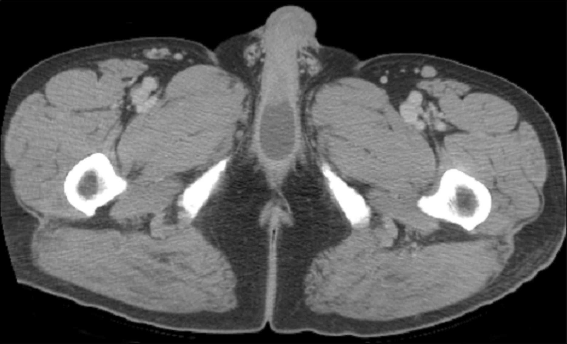 | Fig. 1.The computed tomography scan shows a 4x2.5cm sized well-demarcated cystic mass on the urethra. |
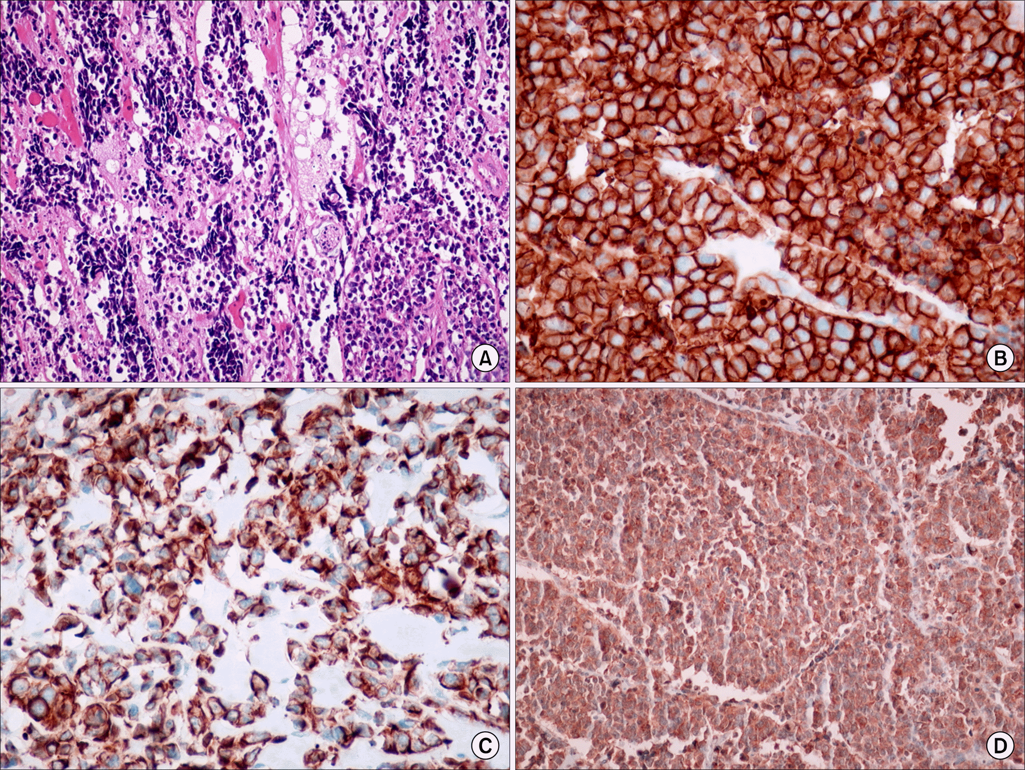 | Fig. 2.(A) The tumor cells display a small, round to oval nucleus with scanty cytoplasm and hyperchromatic nuclei (H&E, x200). (B) The malignant cells show strong positive staining for chromogranin (Immunohistochemistry, x100). (C) The malignant cells show strong positive staining for cytokeratin (Immunohistochemistry, x100). (D) The malignant cells show strong positive staining for synaptophysin (Immunohistochemistry, x100). |
 XML Download