Abstract
Purpose
The aim of this study was to evaluate the effectiveness and indication of radiofrequency ablation (RFA) using renal VX2 tumors by implantation of VX2 tumor cells under the renal capsule in rabbits.
Materials and Methods
Ten rabbits were injected with 30-40μl VX2 tumor cells (1.2x10⁷ viable cells/ml) under the renal capsule of the right kidney by right subcostal incision. On the 14th day after the tumor cells were implanted, we checked for the development of renal tumors, and the sizes and shapes (exophytic or central) of the tumors by the use of computed tomography. We performed RFA in the renal VX2 tumors with a 17G StarBurst electrode through kidney exposure. After the first and third day following RFA, renal function was checked. On the third day, we performed CT and harvested the kidneys for gross and microscopic evaluation.
Results
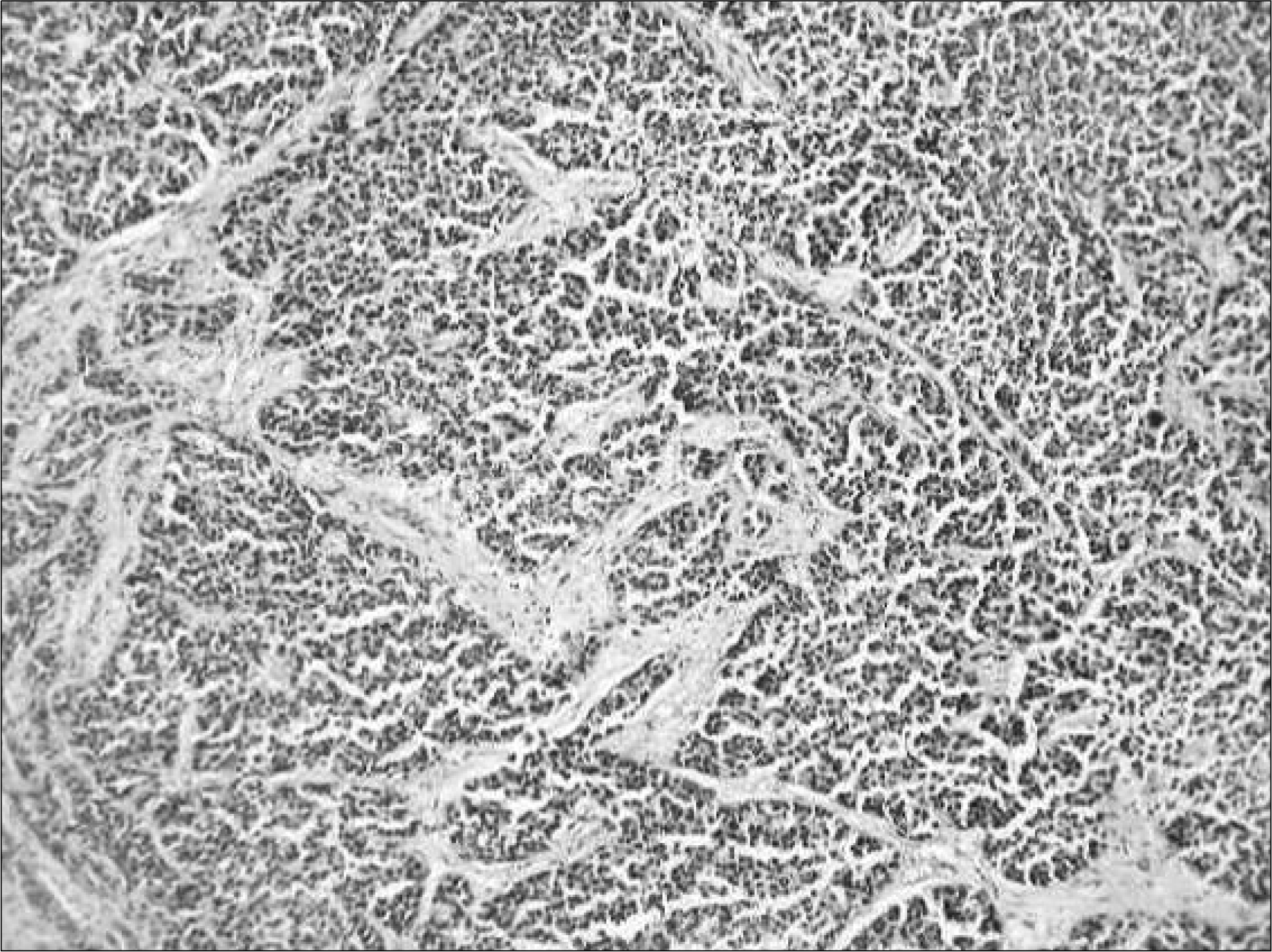
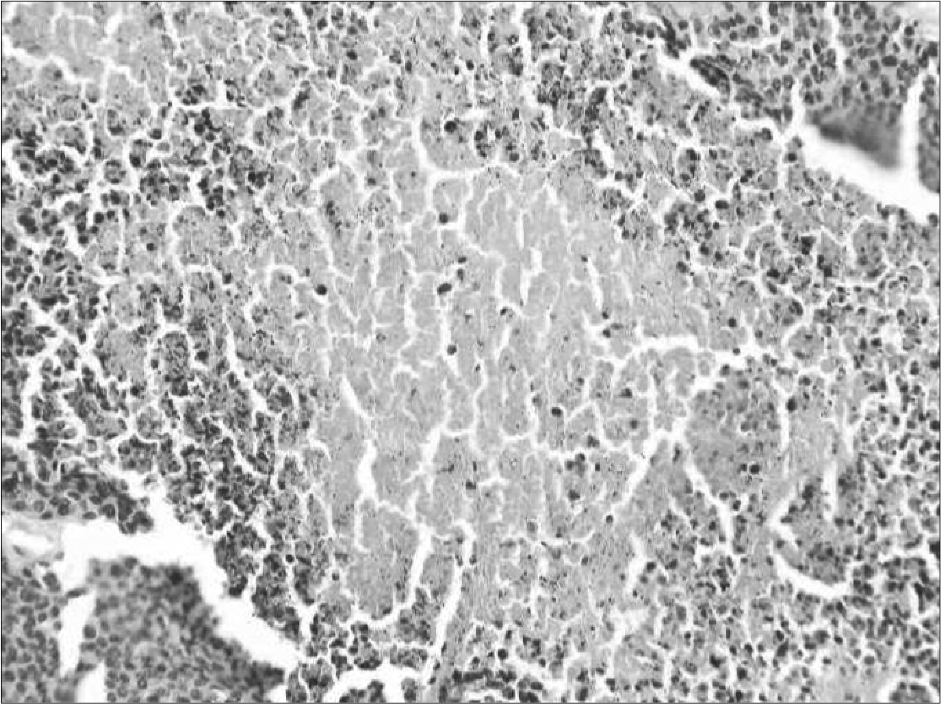
We confirmed the development of renal VX2 tumors in nine cases. Tumor shapes were exophytic in seven cases and central in two cases; the mean size of the tumors was 2.1cm (range, 1.1-3.8cm). In all tumors, RFA was performed. From the use of enhanced CT after RFA on the third day, all of the lesions treated with RFA showed no enhancement. From the pathological findings, coagulative necroses were seen on all of the lesions treated with RFA. The necrotized tumor size after RFA was not different statistically as measured by CT and a pathological examination (p=0.833).
Conclusions
In centrally located renal tumors, we experienced thermal injury in pelvocalyceal systems. RFA is an effective method for nephron sparing surgery as the tumor cells completely disappear and there is preserved renal function and the procedure is easy to apply. We suggest that the RFA method for exophytic renal tumors is more effective than other procedures.
Go to : 
REFERENCES
1. Aslaksen A, Gothlin JH. Imaging of solid renal masses. Curr Opin Radiol. 1991; 3:654–62.
2. Levine E, Huntrakoon M, Wetzel LH. Small renal neoplasms: clinical, pathologic, and imaging features. AJR Am J Roentgenol. 1989; 153:69–73.

3. Uzzo RG, Novick AC. Nephron sparing surgery for renal tumors: indications, techniques and outcomes. J Urol. 2001; 166:6–18.

4. Moll V, Becht E, Ziegler M. Kidney preserving surgery in renal cell tumors: indications, techniques and results in 152 patients. J Urol. 1993; 150:319–23.

5. Lee JW, Jeong IG, Lee ES. Analysis of frequency and predictive factors developing chronic renal diseases after partial or radical nephrectomy about small renal cell carcinoma. Korean J Urol. 2007; 48(Suppl 2):): 104, abstract 112.
6. Kim JW, Ban JH, Jin MH, Yeo JK, Moon DG, Yoon DK. Study of predictive factors about developing renal failure after radical nephrectomy. Korean J Urol. 2007; 48(Suppl 2):): 107, abstract 118.
7. Oakley NE, Hegarty NJ, McNeill A, Gill IS. Minimally invasive nephron-sparing surgery for renal cell cancer. BJU Int. 2006; 98:278–84.

8. Gervais DA, McGovern FJ, Wood BJ, Goldberg SN, McDougal WS, Mueller PR. Radiofrequency ablation of renal cell carcinoma: early clinical experience. Radiology. 2000; 217:665–72.

9. Weizer AZ, Raj GV, O'Connell M, Robertson CN, Nelson RC, Polascik TJ. Complications after percutaneous radiofrequency ablation of renal tumors. Urology. 2005; 66:1176–80.

10. Lee JM, Kim SW, Chung GH, Lee SY, Han YM, Kim CS. Open radiofrequency thermal ablation of renal VX2 tumors in a rabbit model using a cooled-tip electrode: feasibility, safety, and effectiveness. Eur Radiol. 2003; 13:1324–32.

11. Kim SW, Lee JM, Kim CS, Lee SH. Ultrasound-guided radiofrequency thermal ablation of normal kidney in a rabbit model: correlation with CT and histopathology. J Korean Radiol Soc. 2004; 46:25–31.

12. Aron M, Gill IS. Minimally invasive nephron-sparing surgery (MINSS) for renal tumours. Eur Urol. 2007; 51:348–57.

13. Zlotta AR, Wildschutz T, Raviv G, Peny MO, Van Gansbeke D, Noel JC, et al. Radiofrequency interstitial tumor ablation (RITA) is a possible new modality for treatment of renal cancer: ex vivo and in vivo experience. J Endourol. 1997; 11:251–8.

14. Johnson DB, Solomon SB, Su LM, Matsumoto ED, Kavoussi LR, Nakada SY, et al. Defining the complications of cryoablation and radio frequency ablation of small renal tumors: a multi-institutional review. J Urol. 2004; 172:874–7.

15. Pater VR, Leveillee RJ, Hoey MF, Herron AJ, Zaias J, Hulbert JC. Radiofrequency ablation of rabbit kidney using liquid electrode: acute and chronic observations. J Endourol. 2000; 14:155–9.
16. Hafez KS, Fergany AF, Novick AC. Nephron-sparing surgery for localized renal cell carcinoma: impact of tumor size on patient survival, tumor recurrence and TNM staging. J Urol. 1999; 162:1930–3.
17. Curley SA, Izzo F, Delrio P, Ellis LM, Granchi J, Vallone P, et al. Radiofrequency ablation of unresectable primary and metastatic hepatic malignancies: results in 123 patients. Ann Surg. 1999; 230:1–8.
18. Raj GV, Reddan DJ, Hoey MB, Polascik TJ. Management of small renal tumors with radiofrequency ablation. Urology. 2003; 61:23–9.

19. Calkins H, Landberg J, Sousa J, el-Atassi R, Leon A, Kou W, et al. Radiofrequency catheter ablation of accessory atrioventricular connections in 250 patients. Abbreviated therapeutic approach to Wolff-Parkinson-White syndrome. Circulation. 1992; 85:1337–46.

20. Hoey MF, Mulier PM, Leveillee RJ, Hulbert JC. Transurethral prostate ablation with saline electrode allows controlled production of larger lesions than conventional methods. J Endourol. 1997; 11:279–84.

21. Djavan B, Zlotta AR, Susani M, Heinz G, Shariat S, Silverman DE, et al. Transperineal radiofrequency interstitial tumor ablation of the prostate: correlation of magnetic resonance imaging with histopathological examination. Urology. 1997; 50:986–92.
22. Lui KW, Gervais DA, Arellano RA, Mueller PR. Radiofrequency ablation of renal cell carcinoma. Clin Radiol. 2003; 58:905–13.

23. Mahnken AH, Gunther RW, Tacke J. Radiofrequency ablation of renal tumors. Eur Radiol. 2004; 14:1449–55.

24. Su LM, Jarrett TW, Chan DY, Kavoussi LR, Solomon SB. Percutaneous computed tomography-guided radiofrequency ablation of renal masses in high surgical risk patients: preliminary results. Urology. 2003; 61:26–33.

25. Gervais DA, McGovern FJ, Arellano RS, McDougal WS, Mueller PR. Renal cell carcinoma: clinical experience and technical success with radiofrequency ablation of 42 tumors. Radiology. 2003; 226:417–24.

Go to : 
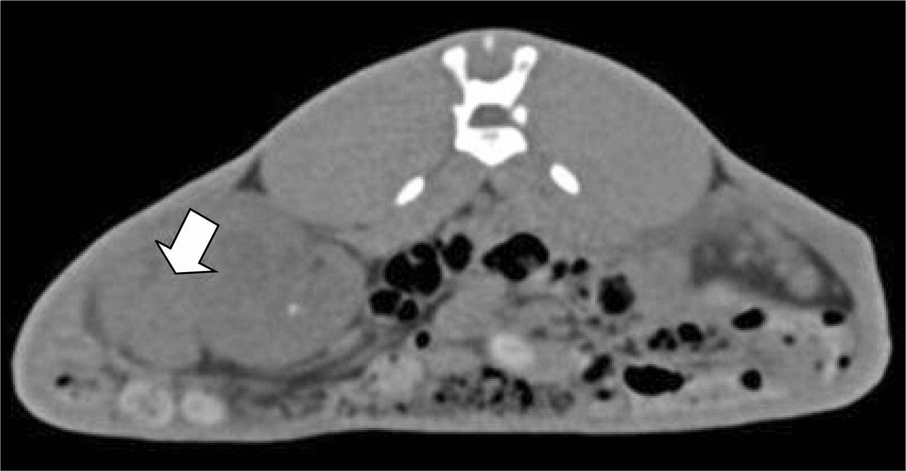 | Fig. 1.A VX2 renal tumor on the right kidney of a rabbit on the 14th day after the implantation of VX2 tumor cells as seen on a pre-enhanced multi-detector computed tomography (MDCT) scan. The tumor shape was exophytic (arrow). |
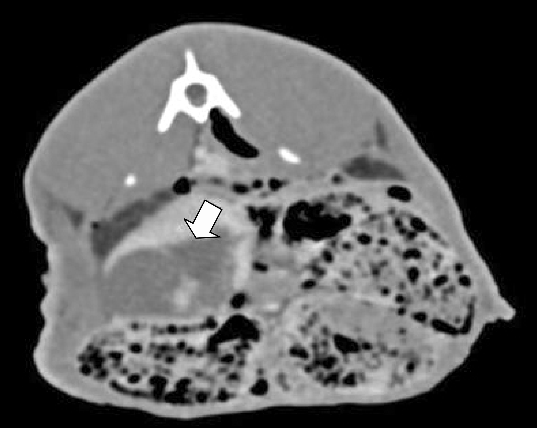 | Fig. 2.On the third day after radiofrequency ablation (RFA) a centrally located lesion of VX2 renal tumor that was treated with RFA was seen as low attenuation in an enhanced multi-detector computed tomography (MDCT) scan (arrow). |
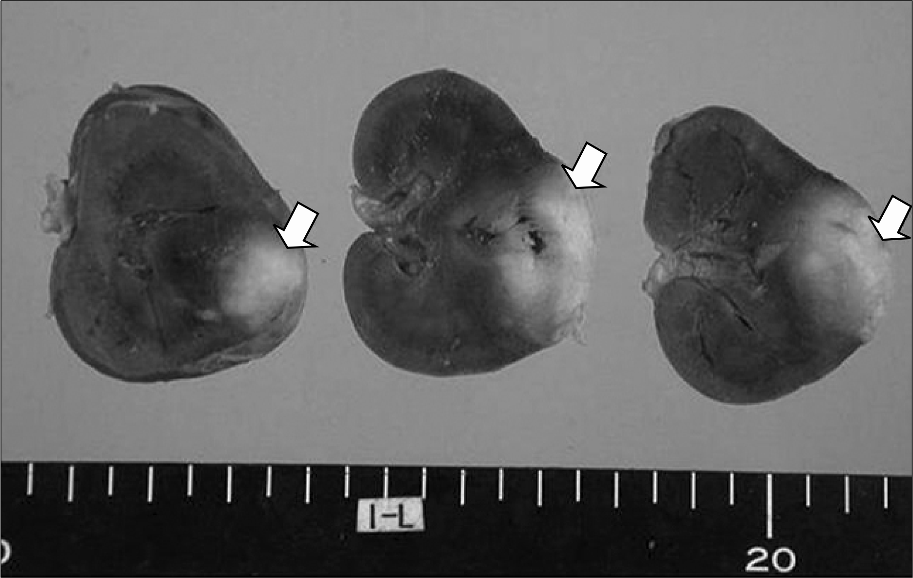 | Fig. 3.Gross findings after radiofrequency ablation (RFA) for an exophytic type renal tumor on the right kidney; tumor necroses were seen (arrow). Pelvocalyceal systems were intact in spite of RFA. |
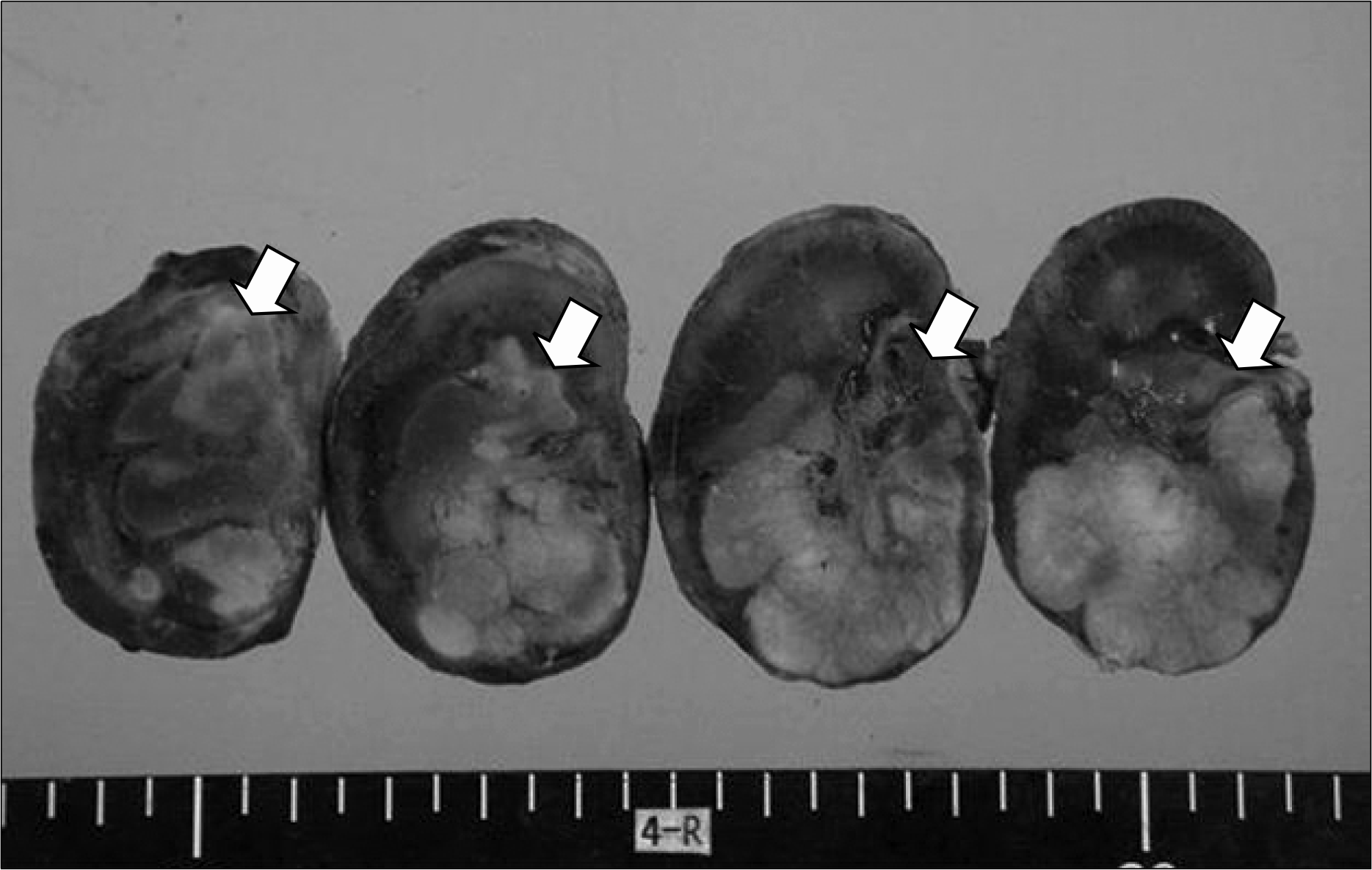 | Fig. 4.Gross findings after radiofrequency ablation (RFA) for a central type renal tumor on the right kidney; tumor necroses were seen (arrow). Pelvocalyceal systems were damaged after RFA. |


 PDF
PDF ePub
ePub Citation
 Print
Print


 XML Download