

 PDF
PDF ePub
ePub Citation
 Print
Print


Abstract
Purpose
Although the purpose of the blood-testis barrier (BTB) is to protect germ cells from harmful influences, it also impedes the delivery of chemotherapeutic agents to the testis. This study was undertaken to determine whether a triolein emulsion could transiently alter the permeability of the BTB in cats.
Materials and Methods
An emulsion of 0.05ml triolein in 20ml of saline or just 20ml of normal saline, as the control, were infused into the testicular arteries in 18 and 15 cats, respectively (embolic and control group). Preand post-contrast magnetic resonance images (MRIs) were obtained 30 minutes and 2 hours after embolization. Qualitative and quantitative analyses of the MRIs were performed via the presence and degree of contrast enhancement and the contrast enhancement ratios (CERs), respectively. An electron microscopy (EM) study was subsequently performed, using a lanthanum tracer, to correlate with the MRI results.
Results
Contrast enhancement of the testis was observed in both groups and at both time points, but was more prominent in the embolic group. The CERs in the embolic group were significantly higher than those in the control group (p=0.0001). In each group, the CERs at 2 hours were significantly lower than those at 30 minutes (p=0.006). In the EM study, the entry of lanthanum was markedly increased at 30 mins, but recovered at 2 hours after embolization compared to the control.
Figures and Tables
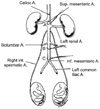 | Fig. 1Schematic drawing showing the abdominal aorta and its main branches. The right and left internal spermatic arteries originating immediately below the renal arteries. Three ligatures (A, B and C) are indicated. A cannula is inserted retrogradely into aorta between the two lower most ligatures, with the tip is pushed beyond the point of the middle ligature (B). During the perfusion procedure, the three ligatures were tied. Ligature A prevented the fixative from reaching the gastrointestinal tract and thorax; ligature B secured the cannula in the aorta, preventing fixative from reaching the iliac vessels and legs; ligature C secured the cannula in the aorta, preventing displacement. Only the portion of the aorta receiving fixative is a 2-3cm region between ligatures A and B; the left renal artery, two internal spermatic (testicular artery) and lumbar vessels originate from this part of the aorta (from Forssmann et al., 1977).
|
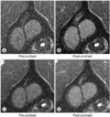 | Fig. 2T1-weighted MR images of the testis at 30 minutes (A and B) and 2 hours (C and D) (TR/TE=320/20) in the control group. At 30 minutes, the testes show mild and diffuse contrast enhancement compared to that of muscle (B). In comparison to the image obtained at 30 minutes (B), the testes show decreased contrast enhancement at 2 hours (D).
|
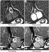 | Fig. 3T1-weighted MR images of the testis at 30 minutes (A and B) and 2 hours (C and D) (TR/TE=320/20) in the embolic group. The testes show marked and homogenous enhancement at 30 minutes (B). In comparison to the image obtained at 30 minutes (B), the testes show decreased contrast enhancement at 2 hours (D).
|
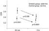 | Fig. 4Plot of the contrast-enhancement ratio (CER) over time. CERs of the embolic group (solid line) are significantly higher than those of the control group (dotted line) at both 30 minutes and 2 hours (p=0.0001). CERs decrease significantly at 2 hours compared to those at 30 minutes (p=0.0006); however, this decrease showed no significant difference between the control and embolic groups (p=0.1889).
|
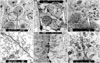 | Fig. 5Morphological electron microscopy study using a lanthanum electron opaque tracer. Upper (A, B and C) and lower (D, E and F) figures are 4,000 and 25,000 times magnified, respectively. Lanthanum was present in the intercellular spaces throughout the seminiferous cord, which can be seen as a fine, dense line (arrow) at 30 mins after the intra-arterial infusion of the triolein emulsion. Compare to the control, the entry of lanthanum was markedly increased at 30 mins, but recovered at 2 hours after the intra-arterial infusion of the triolein emulsion. Lower figures (D, E and F) demonstrate the circle area of the upper figures (A, B and C).
|
References
1. Bart J, Groen HJ, van der Graaf WT, Hollema H, Hendrikse NH, Vaalburg W, et al. An oncological view on the bloodtestis barrier. Lancet Oncol. 2002. 3:357–363.
2. Kamps WA, Bokkerink JP, Hahlen K, Hermans J, Riehm H, Gardner H, et al. Intensive treatment of children with acute lymphoblastic leukemia according to ALL-BFM-86 without cranial radiotherapy: results of Dutch Childhood Leukemia Study Group Protocol ALL-7 (1988-1991). Blood. 1999. 94:1226–1236.
3. Miniero R, Saracco P, Pastore G, Zurlo MG, Terracini B, Rosso P, et al. Relapse after first cessation of therapy in childhood acute lymphoblastic leukemia: a 10-year follow-up study. Italian Association of Pediatric Hematology-Oncology (AIEOP). Med Pediatr Oncol. 1995. 24:71–76.
4. Touroutoglou N, Dimopoulos MA, Younes A, Hess M, Pugh W, Cox J, et al. Testicular lymphoma: late relapses and poor outcome despite doxorubicin-based therapy. J Clin Oncol. 1995. 13:1361–1367.
5. Tondini C, Ferreri AJ, Siracusano L, Valagussa P, Giardini R, Rampinelli I, et al. Diffuse large-cell lymphoma of the testis. J Clin Oncol. 1999. 17:2854–2858.
6. Kroll RA, Neuwelt EA. Outwitting the blood-brain barrier for therapeutic purposes: osmotic opening and other means. Neurosurgery. 1998. 42:1083–1099.
7. Kim HJ, Lee CH, Kim HG, Lee SD, Son SM, Kim YW, et al. Reversible MR changes in the cat brain after cerebral fat embolism induced by triolein emulsion. AJNR Am J Neuroradiol. 2004. 25:958–963.
8. Sun EL, Gondos B. Formation of the blood-testis barrier in the rabbit. Cell Tissue Res. 1986. 243:575–578.
9. Forssmann WG, Ito S, Weihe E, Aoki A, Dym M, Fawcett DW. An improved perfusion fixation method for the testis. Anat Rec. 1977. 188:307–314.
10. Runge VM, Price AC, Wehr CJ, Atkinson JB, Tweedle MF. Contrast enhanced MRI: evaluation of a canine model of osmotic blood-brain barrier disruption. Invest Radiol. 1985. 20:830–844.
11. Farghali H, Williams DS, Simplaceanu E, Ho C, Van Thiel DH. An evaluation of the integrity of the blood-testis barrier by magnetic resonance imaging. Magn Reson Med. 1991. 22:81–87.
12. Kim HJ, Lee CH, Lee SH, Moon TY. Early development of vasogenic edema in experimental cerebral fat embolism in cat. Invest Radiol. 2001. 36:460–469.
13. Eng F, Wiebe JP, Alima LH. Long-term alterations in the permeability of the blood-testis barrier following a single intratesticular injection of dilute aqueous glycerol. J Androl. 1994. 15:311–317.
14. Wiebe JP, Kowalik A, Gallardi RL, Egeler O, Clubb BH. Glycerol disrupts tight junction-associated actin microfilaments, occludin, and microtubules in Sertoli cells. J Androl. 2000. 21:625–635.
15. Dym M, Fawcett DW. The blood-testis barrier in the rat and the physiological compartment of the seminiferous epithelium. Biol Reprod. 1970. 3:308–326.
 XML Download