

 PDF
PDF ePub
ePub Citation
 Print
Print


Abstract
There has been only 23 cases of neonatal adrenal hemorrhage presenting as an acute scrotum and unnecessary surgical exploration was performed in nine of these case (39%) due to suspected testicular torsion. We report here on a case of a 2-day-old boy with neonatal adrenal hemorrhage, and he presented with an acute scrotum; this child's condition was managed conservatively.
References
1. Black J, Williams DI. Natural history of adrenal hemorrhage in the newborn. Arch Dis Child. 1973; 48:183–90.
2. Adorisio O, Mattei R, Ciardini E, Centonze N, Noccioli B. Neonatal adrenal hemorrhage mimicking an acute scrotum. J Perinatol. 2007; 27:130–2.

3. Avolio L, Fusillo M, Ferrari G, Chiara A, Bragheri R. Neonatal adrenal hemorrhage manifesting as acute scrotum: timely diagnosis prevents unnecessary surgery. Urology. 2002; 59:601.
4. Giacoia GP, Cravens JD. Neonatal adrenal hemorrhage presenting as scrotal hematoma. J Urol. 1990; 143:567–8.
5. Karpe B, Nybonde T. Adrenal hemorrhage versus testicular torsion-a diagnostic dilemma in the neonate. Pediatr Surg Int. 1989; 4:337–40.
6. Putnam MH. Neonatal adrenal hemorrhage presenting as a right scrotal mass. JAMA. 1989; 261:2958.
7. Amoury RA, Barth GW, Hall RT, Rhodes PG, Holder TM, Ashcraft KW. Scrotal ecchymosis: sign of intraperitoneal hemorrhage in the newborn South Med J. 1982; 75:1471–5.
8. Choo GY, Chung YG, Kim YJ, Chung JS, Kang SC, Lee T. Bilateral neonatal torsion of testis. Korean J Urol. 2006; 47:794–6.
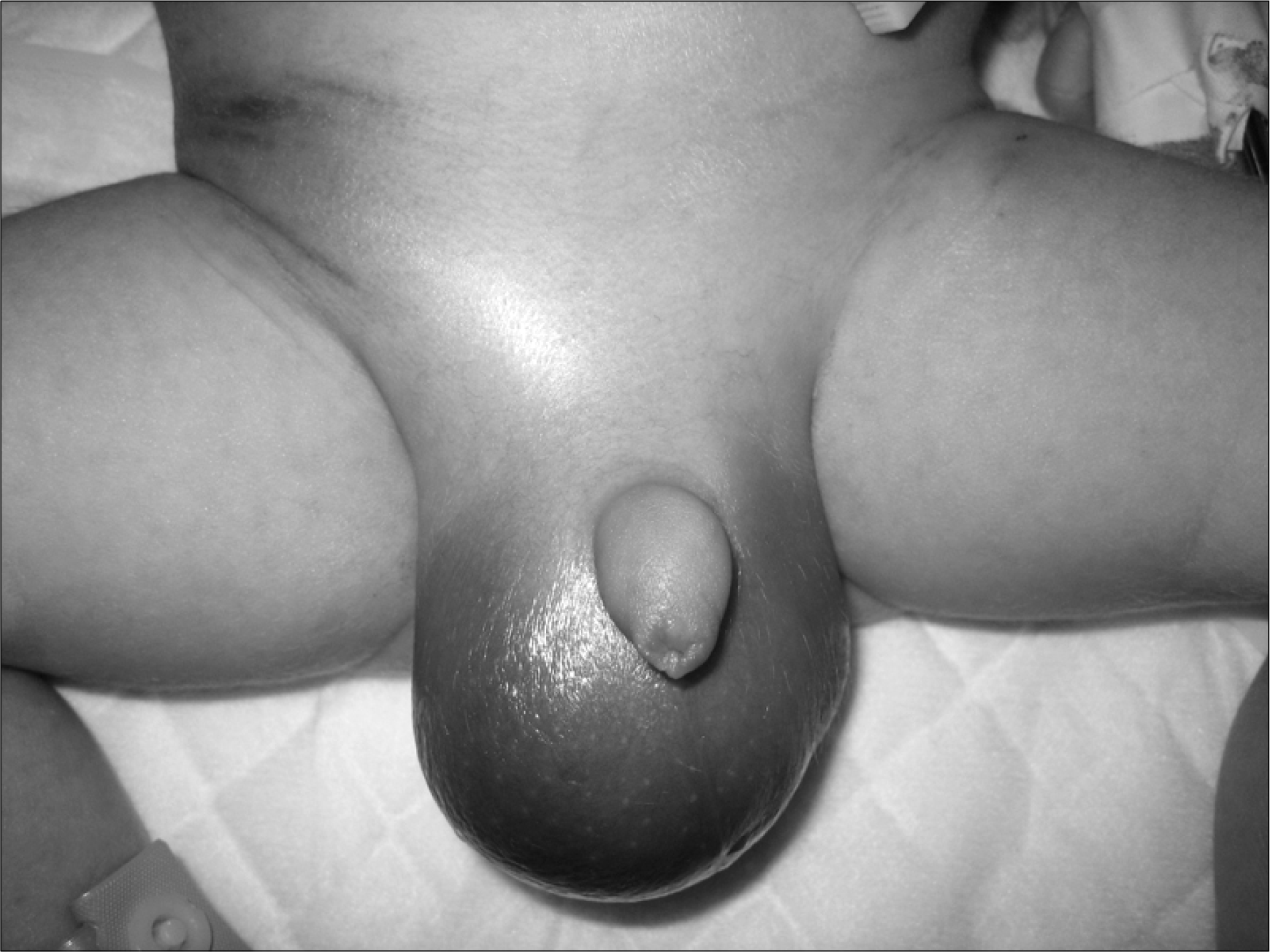
Fig. 2.
The scrotal (A) and transabdominal (B) ultrasonography demonstrated a right testis (T) with surrounding hematoma (H) and a retroperitoneal hematoma (R) above the right kidney (K).

Fig. 3.
Magnetic resonance imaging demonstrated a retroperitoneal hematoma (H) with low, heterogeneous signal intensity on the T2-weighted image (A), and this was not enhanced on the contrast enhanced T1-weighted image (B).
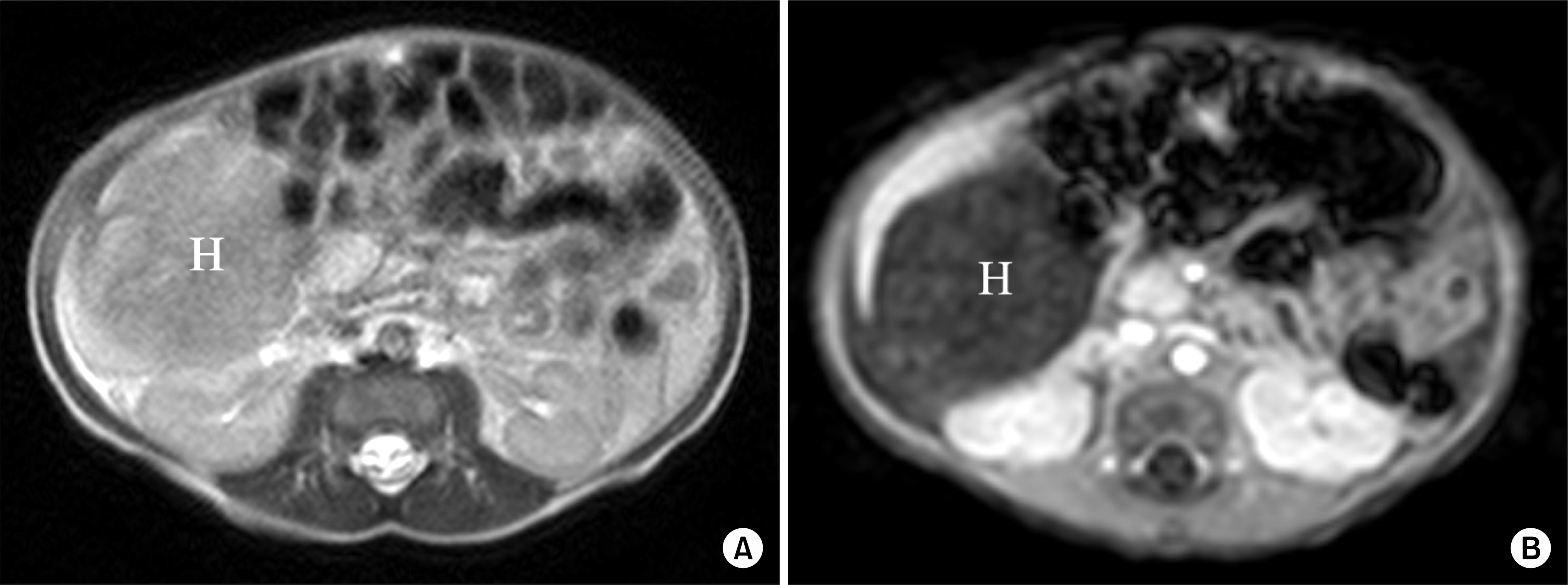
Table 1.
The reported cases of neonatal adrenal hemorrhage presenting as acute scrotum: clinical characteristics of the patients
 XML Download